4MX3

Crystal Structure of PKA RIalpha Homodimer


Experimental Data Snapshot

  • Method: X-RAY DIFFRACTION
  • Resolution: 3.88 Å
  • R-Value Free: 0.287 
  • R-Value Work: 0.260 
  • R-Value Observed: 0.261 

wwPDB Validation   3D Report Full Report


Ligand Structure Quality Assessment 


This is version 1.2 of the entry. See complete history


Literature

PKA RI alpha Homodimer Structure Reveals an Intermolecular Interface with Implications for Cooperative cAMP Binding and Carney Complex Disease.

Bruystens, J.G.Wu, J.Fortezzo, A.Kornev, A.P.Blumenthal, D.K.Taylor, S.S.

(2014) Structure 22: 59-69

  • DOI: https://doi.org/10.1016/j.str.2013.10.012
  • Primary Citation of Related Structures:  
    4MX3

  • PubMed Abstract: 

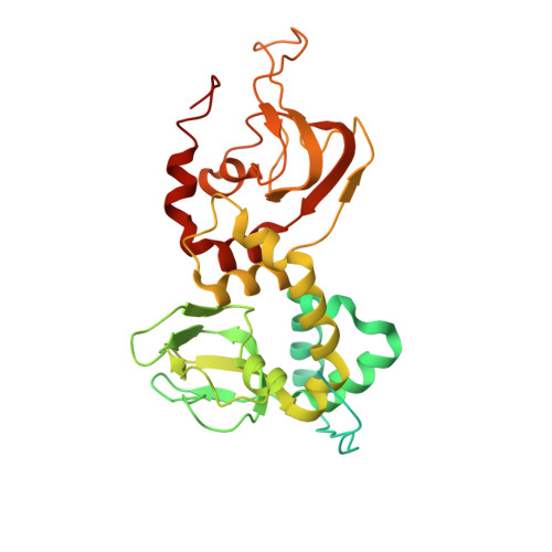
    The regulatory (R) subunit is the cAMP receptor of protein kinase A. Following cAMP binding, the inactive PKA holoenzyme complex separates into two active catalytic (C) subunits and a cAMP-bound R dimer. Thus far, only monomeric R structures have been solved, which fell short in explaining differences of cAMP binding for the full-length protein as compared to the truncated R subunits. Here we solved a full-length R-dimer structure that reflects the biologically relevant conformation, and this structure agrees well with small angle X-ray scattering. An isoform-specific interface is revealed between the protomers. This interface acts as an intermolecular sensor for cAMP and explains the cooperative character of cAMP binding to the RIα dimer. Mutagenesis of residues on this interface not only leads to structural and biochemical changes, but is also linked to Carney complex disease.


  • Organizational Affiliation

    Department of Chemistry and Biochemistry, University of California at San Diego, La Jolla, CA 92093, USA.


Macromolecules
Find similar proteins by:  (by identity cutoff)  |  3D Structure
Entity ID: 1
MoleculeChains Sequence LengthOrganismDetailsImage
cAMP-dependent protein kinase type I-alpha regulatory subunit
A, B
379Bos taurusMutation(s): 0 
Gene Names: PRKAR1A
UniProt
Find proteins for P00514 (Bos taurus)
Explore P00514 
Go to UniProtKB:  P00514
Entity Groups  
Sequence Clusters30% Identity50% Identity70% Identity90% Identity95% Identity100% Identity
UniProt GroupP00514
Sequence Annotations
Expand
  • Reference Sequence
Experimental Data & Validation

Experimental Data

  • Method: X-RAY DIFFRACTION
  • Resolution: 3.88 Å
  • R-Value Free: 0.287 
  • R-Value Work: 0.260 
  • R-Value Observed: 0.261 
  • Space Group: P 41 21 2
Unit Cell:
Length ( Å )Angle ( ˚ )
a = 104.672α = 90
b = 104.672β = 90
c = 218.327γ = 90
Software Package:
Software NamePurpose
ADSCdata collection
PHASESphasing
REFMACrefinement
HKL-2000data reduction
HKL-2000data scaling

Structure Validation

View Full Validation Report


Ligand Structure Quality Assessment 


Entry History 

Deposition Data

Revision History  (Full details and data files)

  • Version 1.0: 2014-01-15
    Type: Initial release
  • Version 1.1: 2014-02-05
    Changes: Database references
  • Version 1.2: 2023-09-20
    Changes: Data collection, Database references, Derived calculations, Refinement description