4MWI

Crystal structure of the human MLKL pseudokinase domain


Experimental Data Snapshot

  • Method: X-RAY DIFFRACTION
  • Resolution: 1.70 Å
  • R-Value Free: 0.194 
  • R-Value Work: 0.165 
  • R-Value Observed: 0.166 

wwPDB Validation   3D Report Full Report


Ligand Structure Quality Assessment 


This is version 1.3 of the entry. See complete history


Literature

Insights into the evolution of divergent nucleotide-binding mechanisms among pseudokinases revealed by crystal structures of human and mouse MLKL.

Murphy, J.M.Lucet, I.S.Hildebrand, J.M.Tanzer, M.C.Young, S.N.Sharma, P.Lessene, G.Alexander, W.S.Babon, J.J.Silke, J.Czabotar, P.E.

(2014) Biochem J 457: 369-377

  • DOI: https://doi.org/10.1042/BJ20131270
  • Primary Citation of Related Structures:  
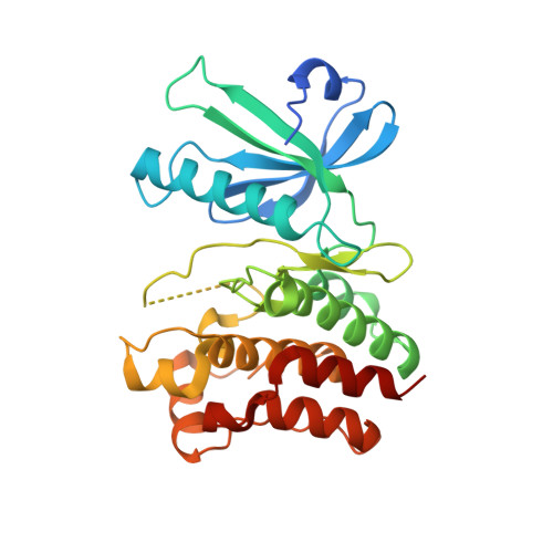
    4MWI

  • PubMed Abstract: 

    The pseudokinase MLKL (mixed lineage kinase domain-like) was identified recently as an essential checkpoint in the programmed necrosis or 'necroptosis' cell death pathway. In the present study, we report the crystal structure of the human MLKL pseudokinase domain at 1.7 Å (1 Å=0.1 nm) resolution and probe its nucleotide-binding mechanism by performing structure-based mutagenesis. By comparing the structures and nucleotide-binding determinants of human and mouse MLKL orthologues, the present study provides insights into the evolution of nucleotide-binding mechanisms among pseudokinases and their mechanistic divergence from conventional catalytically active protein kinases.


  • Organizational Affiliation

    ‡Department of Biochemistry and Molecular Biology, School of Biomedical Sciences, Monash University, Clayton, Victoria 3800, Australia.


Macromolecules
Find similar proteins by:  (by identity cutoff)  |  3D Structure
Entity ID: 1
MoleculeChains Sequence LengthOrganismDetailsImage
Mixed lineage kinase domain-like protein294Homo sapiensMutation(s): 0 
Gene Names: MLKL
UniProt & NIH Common Fund Data Resources
Find proteins for Q8NB16 (Homo sapiens)
Explore Q8NB16 
Go to UniProtKB:  Q8NB16
PHAROS:  Q8NB16
GTEx:  ENSG00000168404 
Entity Groups  
Sequence Clusters30% Identity50% Identity70% Identity90% Identity95% Identity100% Identity
UniProt GroupQ8NB16
Sequence Annotations
Expand
  • Reference Sequence
Small Molecules
Ligands 2 Unique
IDChains Name / Formula / InChI Key2D Diagram3D Interactions
PXN
Query on PXN

Download Ideal Coordinates CCD File 
B [auth A](2S)-1-[3-{[(2R)-2-hydroxypropyl]oxy}-2,2-bis({[(2R)-2-hydroxypropyl]oxy}methyl)propoxy]propan-2-ol
C17 H36 O8
GXEZGLLPFFKHGE-FPCVCCKLSA-N
GOL
Query on GOL

Download Ideal Coordinates CCD File 
C [auth A]GLYCEROL
C3 H8 O3
PEDCQBHIVMGVHV-UHFFFAOYSA-N
Experimental Data & Validation

Experimental Data

  • Method: X-RAY DIFFRACTION
  • Resolution: 1.70 Å
  • R-Value Free: 0.194 
  • R-Value Work: 0.165 
  • R-Value Observed: 0.166 
  • Space Group: C 2 2 21
Unit Cell:
Length ( Å )Angle ( ˚ )
a = 72.114α = 90
b = 74.723β = 90
c = 127.601γ = 90
Software Package:
Software NamePurpose
Blu-Icedata collection
PHASERphasing
PHENIXrefinement
XDSdata reduction
XDSdata scaling

Structure Validation

View Full Validation Report


Ligand Structure Quality Assessment 


Entry History 

Deposition Data

Revision History  (Full details and data files)

  • Version 1.0: 2013-12-04
    Type: Initial release
  • Version 1.1: 2014-03-19
    Changes: Database references
  • Version 1.2: 2014-04-16
    Changes: Other
  • Version 1.3: 2023-09-20
    Changes: Data collection, Database references, Derived calculations, Refinement description