The multiple nucleotide-divalent cation binding modes of Saccharomyces cerevisiae CK2 alpha indicate a possible co-substrate hydrolysis product (ADP/GDP) release pathway.
Liu, H., Wang, H., Teng, M., Li, X.(2014) Acta Crystallogr D Biol Crystallogr 70: 501-513
- PubMed: 24531484
- DOI: https://doi.org/10.1107/S1399004713027879
- Primary Citation of Related Structures:
4JQE, 4JR7, 4LFI, 4MWH - PubMed Abstract:
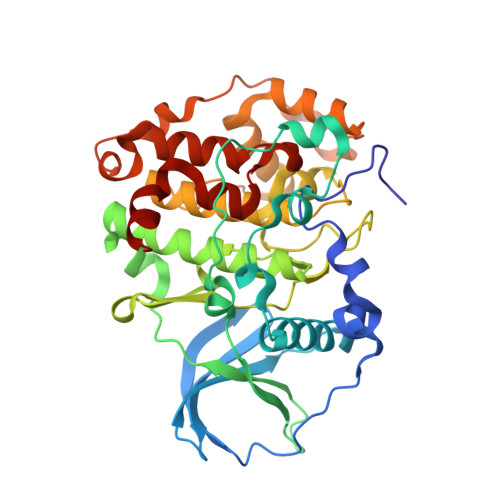
CK2 is a ubiquitous and conserved protein kinase in eukaryotic organisms and is important in many biological processes. It is unique in maintaining constitutive activity and in using both ATP and GTP as phosphor donors. In this study, crystal structures of recombinant Saccharomyces cerevisiae CK2α (scCK2α) complexed with GMPPNP, ATP and AMPPN with either Mg2+ or Mn2+ as the coordinated divalent cation are presented. The overall structure of scCK2α shows high similarity to its homologous proteins by consisting of two domains with the co-substrate lying in the cleft between them. However, three characteristic features distinguish scCK2α from its homologues. Firstly, the Lys45-Glu53 and Arg48-Glu53 interactions in scCK2α lead Lys50 to adopt a unique conformation that is able to stabilize the γ-phosphate of the co-substrate, which makes the existence of the `essential divalent cation' not so essential. The multiple nucleotide-divalent cation binding modes of the active site of scCK2α are apparently different from the two-divalent-cation-occupied active site of Zea mays CK2α and human CK2α. Secondly, conformational change of Glu53 in scCK2α-AMPPN breaks its interaction with Lys45 and Arg48; as a result, the co-substrate binding pocket becomes more open. This may suggest a clue to a possible ADP/GDP-release pathway, because the NE1 atom of the Trp in the `DWG motif' of CK2α forms a hydrogen bond to the O atom of Leu212, which seems to make ADP release by means of the `DFG-in flip to DFG-out' model found in most eukaryotic protein kinases impossible. Coincidentally, two sulfate ions which may mimic two phosphate groups were captured by Arg161 and Lys197 around the pocket. Mutagenesis and biochemical experiments on R161A and K197A mutants support the above proposal. Finally, scCK2α is unique in containing an insertion region whose function had not been identified in previous research. It is found that the insertion region contributes to maintaining the constitutively active conformation of the scCK2α catalytic site, but does not participate in interaction with the regulatory subunits.
Organizational Affiliation:
School of Life Sciences, University of Science and Technology of China, 96 Jinzhai Road, Hefei, Anhui 230026, People's Republic of China.