Implementing Fluorescence Anisotropy Screening and Crystallographic Analysis to Define PKA Isoform-Selective Activation by cAMP Analogs.
Brown, S.H., Cheng, C.Y., Saldanha, S.A., Wu, J., Cottam, H.B., Sankaran, B., Taylor, S.S.(2013) ACS Chem Biol 8: 2164-2172
- PubMed: 23978166
- DOI: https://doi.org/10.1021/cb400247t
- Primary Citation of Related Structures:
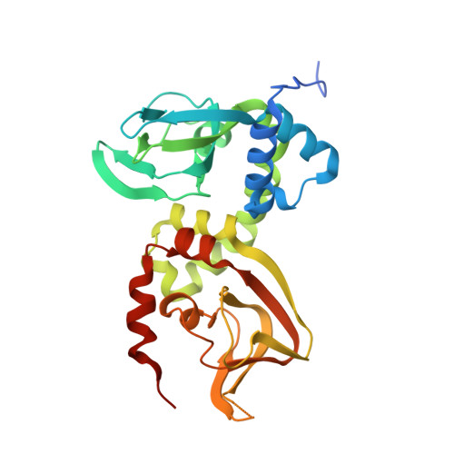
4JV4, 4JVA - PubMed Abstract:
Cyclic AMP (cAMP) is a ubiquitous second messenger that regulates many proteins, most notably cAMP-dependent protein kinase (PKA). PKA holoenzymes (comprised of two catalytic (C) and two regulatory (R) subunits) regulate a wide variety of cellular processes, and its functional diversity is amplified by the presence of four R-subunit isoforms, RIα, RIβ, RIIα, and RIIβ. Although these isoforms all respond to cAMP, they are functionally nonredundant and exhibit different biochemical properties. In order to understand the functional differences between these isoforms, we screened cAMP derivatives for their ability to selectively activate RI and RII PKA holoenzymes using a fluorescence anisotropy assay. Our results indicate that RIα holoenzymes are selectively activated by C8-substituted analogs and RIIβ holoenzymes by N6-substituted analogs, where HE33 is the most prominent RII activator. We also solved the crystal structures of both RIα and RIIβ bound to HE33. The RIIβ structure shows the bulky aliphatic substituent of HE33 is fully encompassed by a pocket comprising of hydrophobic residues. RIα lacks this hydrophobic lining in Domain A, and the side chains are displaced to accommodate the HE33 dipropyl groups. Comparison between cAMP-bound structures reveals that RIIβ, but not RIα, contains a cavity near the N6 site. This study suggests that the selective activation of RII over RI isoforms by N6 analogs is driven by the spatial and chemical constraints of Domain A and paves the way for the development of potent noncyclic nucleotide activators to specifically target PKA iso-holoenyzmes.
Organizational Affiliation:
Department of Chemistry and Biochemistry, ‡Department of Pharmacology and Howard Hughes Medical Institute, §Moores Cancer Center, University of California , San Diego, La Jolla, California 92037-0654, United States.