4JLC

Crystal structure of mouse TBK1 bound to SU6668


Experimental Data Snapshot

  • Method: X-RAY DIFFRACTION
  • Resolution: 3.00 Å
  • R-Value Free: 0.267 
  • R-Value Work: 0.234 
  • R-Value Observed: 0.237 

wwPDB Validation   3D Report Full Report


Ligand Structure Quality Assessment 


This is version 1.2 of the entry. See complete history


Literature

Structural Insights into the Functions of TBK1 in Innate Antimicrobial Immunity.

Shu, C.Sankaran, B.Chaton, C.T.Herr, A.B.Mishra, A.Peng, J.Li, P.

(2013) Structure 21: 1137-1148

  • DOI: https://doi.org/10.1016/j.str.2013.04.025
  • Primary Citation of Related Structures:  
    4JL9, 4JLC

  • PubMed Abstract: 

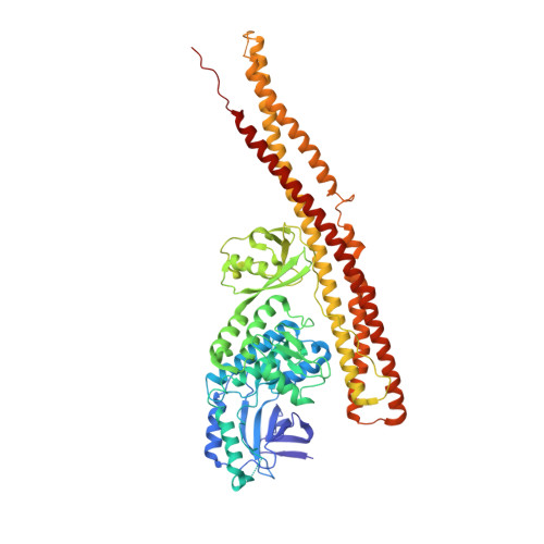
    Tank-binding kinase 1 (TBK1) is a serine/threonine protein-kinase mediating innate antimicrobial immunity. TBK1 is involved in the signaling of TLRs, RLRs, and STING-mediated sensing of cytosolic DNA. Stimulation of these receptors results in the activation of TBK1, which phosphorylates interferon regulatory factor (IRF)-3. Phosphorylated IRF-3 translocates into the nucleus to initiate the transcription of the interferon (IFN)-β gene. Here, we show that TBK1 is activated by autophosphorylation at residue Ser172. Structures of TBK1 bound to two inhibitors showed that TBK1 has the IκB kinase fold with three distinct domains: the kinase domain, the ubiquitin-like domain, and the scaffold and dimerization domain. However, the overall structures of the TBK1 monomer and its dimer are different from IKKβ in the arrangements of the three domains and in dimer formation. Phosphorylation of IRF-3 by TBK1 in vitro results in its oligomerization, and phosphorylation of residue Ser386 plays a key role in IRF-3 activation.


  • Organizational Affiliation

    Department of Biochemistry and Biophysics, Texas A&M University, College Station, TX 77843-2128, USA.


Macromolecules
Find similar proteins by:  (by identity cutoff)  |  3D Structure
Entity ID: 1
MoleculeChains Sequence LengthOrganismDetailsImage
Serine/threonine-protein kinase TBK1657Mus musculusMutation(s): 0 
Gene Names: Tbk1
EC: 2.7.11.1
UniProt
Find proteins for Q9WUN2 (Mus musculus)
Explore Q9WUN2 
Go to UniProtKB:  Q9WUN2
Entity Groups  
Sequence Clusters30% Identity50% Identity70% Identity90% Identity95% Identity100% Identity
UniProt GroupQ9WUN2
Sequence Annotations
Expand
  • Reference Sequence
Small Molecules
Ligands 1 Unique
IDChains Name / Formula / InChI Key2D Diagram3D Interactions
SU6
Query on SU6

Download Ideal Coordinates CCD File 
B [auth A]3-{2,4-dimethyl-5-[(Z)-(2-oxo-1,2-dihydro-3H-indol-3-ylidene)methyl]-1H-pyrrol-3-yl}propanoic acid
C18 H18 N2 O3
NHFDRBXTEDBWCZ-ZROIWOOFSA-N
Experimental Data & Validation

Experimental Data

  • Method: X-RAY DIFFRACTION
  • Resolution: 3.00 Å
  • R-Value Free: 0.267 
  • R-Value Work: 0.234 
  • R-Value Observed: 0.237 
  • Space Group: P 32 2 1
Unit Cell:
Length ( Å )Angle ( ˚ )
a = 140.48α = 90
b = 140.48β = 90
c = 86.56γ = 120
Software Package:
Software NamePurpose
ADSCdata collection
MOLREPphasing
PHENIXrefinement
MOSFLMdata reduction
SCALAdata scaling

Structure Validation

View Full Validation Report


Ligand Structure Quality Assessment 


Entry History 

Deposition Data

  • Released Date: 2013-06-19 
  • Deposition Author(s): Li, P.

Revision History  (Full details and data files)

  • Version 1.0: 2013-06-19
    Type: Initial release
  • Version 1.1: 2013-07-17
    Changes: Database references
  • Version 1.2: 2023-09-20
    Changes: Data collection, Database references, Derived calculations, Refinement description