4J8Z

Crystal Structure of the Human SPOP BTB Domain


Experimental Data Snapshot

  • Method: X-RAY DIFFRACTION
  • Resolution: 2.42 Å
  • R-Value Free: 0.255 
  • R-Value Work: 0.207 
  • R-Value Observed: 0.209 

wwPDB Validation   3D Report Full Report


This is version 1.2 of the entry. See complete history


Literature

Structural basis of high-order oligomerization of the cullin-3 adaptor SPOP

Geersdaele, L.K.Stead, M.A.Harrison, C.M.Carr, S.B.Close, H.J.Rosbrook, G.O.Connell, S.D.Wright, S.C.

(2013) Acta Crystallogr D Biol Crystallogr 69: 1677-1684

  • DOI: https://doi.org/10.1107/S0907444913012687
  • Primary Citation of Related Structures:  
    4HS2, 4J8Z

  • PubMed Abstract: 

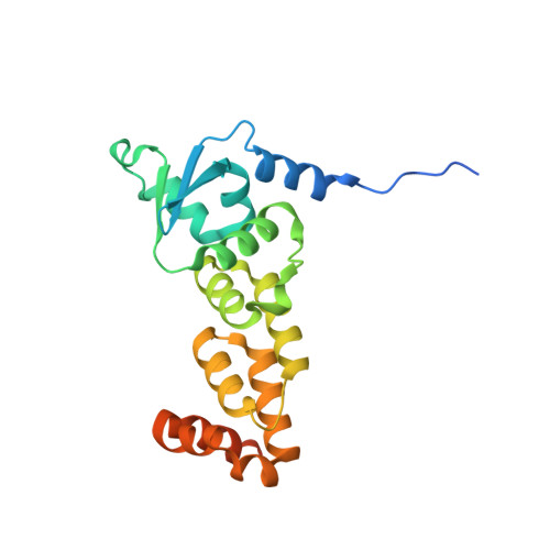
    Protein ubiquitination in eukaryotic cells is mediated by diverse E3 ligase enzymes that each target specific substrates. The cullin E3 ligase complexes are the most abundant class of E3 ligases; they contain various cullin components that serve as scaffolds for interaction with substrate-recruiting adaptor proteins. SPOP is a BTB-domain adaptor of the cullin-3 E3 ligase complexes; it selectively recruits substrates via its N-terminal MATH domain, whereas its BTB domain mediates dimerization and interactions with cullin-3. It has recently been recognized that the high-order oligomerization of SPOP enhances the ubiquitination of substrates. Here, a dimerization interface in the SPOP C-terminus is identified and it is shown that the dimerization interfaces of the BTB domain and of the C-terminus act independently and in tandem to generate high-order SPOP oligomers. The crystal structure of the dimeric SPOP C-terminal domain is reported at 1.5 Å resolution and it is shown that Tyr353 plays a critical role in high-order oligomerization. A model of the high-order SPOP oligomer is presented that depicts a helical organization that could enhance the efficiency of substrate ubiquitination.


  • Organizational Affiliation

    School of Biology, University of Leeds, Leeds LS2 9JT, England.


Macromolecules
Find similar proteins by:  (by identity cutoff)  |  3D Structure
Entity ID: 1
MoleculeChains Sequence LengthOrganismDetailsImage
Speckle-type POZ protein
A, B
211Homo sapiensMutation(s): 1 
Gene Names: SPOP
UniProt & NIH Common Fund Data Resources
Find proteins for O43791 (Homo sapiens)
Explore O43791 
Go to UniProtKB:  O43791
PHAROS:  O43791
GTEx:  ENSG00000121067 
Entity Groups  
Sequence Clusters30% Identity50% Identity70% Identity90% Identity95% Identity100% Identity
UniProt GroupO43791
Sequence Annotations
Expand
  • Reference Sequence
Experimental Data & Validation

Experimental Data

  • Method: X-RAY DIFFRACTION
  • Resolution: 2.42 Å
  • R-Value Free: 0.255 
  • R-Value Work: 0.207 
  • R-Value Observed: 0.209 
  • Space Group: C 1 2 1
Unit Cell:
Length ( Å )Angle ( ˚ )
a = 111.39α = 90
b = 71.58β = 117.21
c = 62.22γ = 90
Software Package:
Software NamePurpose
SCALAdata scaling
PHASERphasing
REFMACrefinement
PDB_EXTRACTdata extraction
XDSdata scaling
XDSdata reduction
Aimlessdata scaling

Structure Validation

View Full Validation Report


Entry History 

Deposition Data

Revision History  (Full details and data files)

  • Version 1.0: 2013-09-11
    Type: Initial release
  • Version 1.1: 2014-01-22
    Changes: Database references
  • Version 1.2: 2024-03-20
    Changes: Data collection, Database references, Refinement description