4GL9

Crystal structure of inhibitory protein SOCS3 in complex with JAK2 kinase domain and fragment of GP130 intracellular domain


Experimental Data Snapshot

  • Method: X-RAY DIFFRACTION
  • Resolution: 3.90 Å
  • R-Value Free: 0.281 
  • R-Value Work: 0.249 
  • R-Value Observed: 0.254 

wwPDB Validation   3D Report Full Report


Ligand Structure Quality Assessment 


This is version 1.4 of the entry. See complete history


Literature

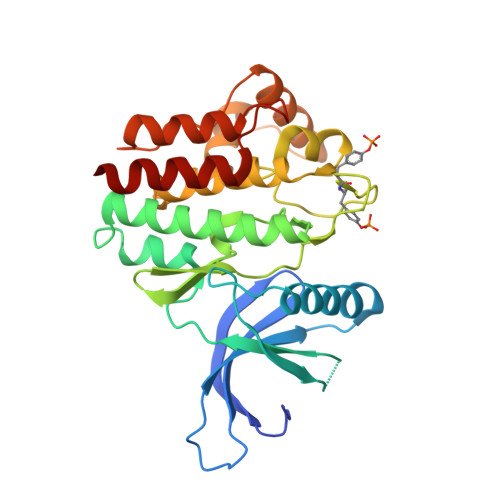
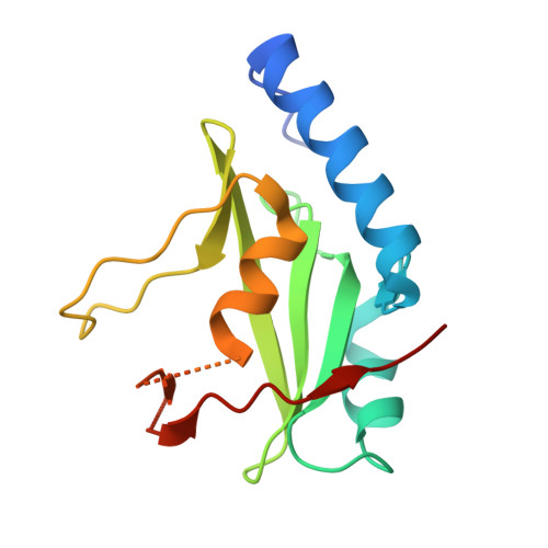
SOCS3 binds specific receptor-JAK complexes to control cytokine signaling by direct kinase inhibition.

Kershaw, N.J.Murphy, J.M.Liau, N.P.Varghese, L.N.Laktyushin, A.Whitlock, E.L.Lucet, I.S.Nicola, N.A.Babon, J.J.

(2013) Nat Struct Mol Biol 20: 469-476

  • DOI: https://doi.org/10.1038/nsmb.2519
  • Primary Citation of Related Structures:  
    4GL9

  • PubMed Abstract: 

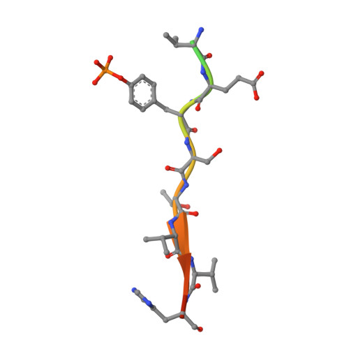
    The inhibitory protein SOCS3 plays a key part in the immune and hematopoietic systems by regulating signaling induced by specific cytokines. SOCS3 functions by inhibiting the catalytic activity of Janus kinases (JAKs) that initiate signaling within the cell. We determined the crystal structure of a ternary complex between mouse SOCS3, JAK2 (kinase domain) and a fragment of the interleukin-6 receptor β-chain. The structure shows that SOCS3 binds JAK2 and receptor simultaneously, using two opposing surfaces. While the phosphotyrosine-binding groove on the SOCS3 SH2 domain is occupied by receptor, JAK2 binds in a phosphoindependent manner to a noncanonical surface. The kinase-inhibitory region of SOCS3 occludes the substrate-binding groove on JAK2, and biochemical studies show that it blocks substrate association. These studies reveal that SOCS3 targets specific JAK-cytokine receptor pairs and explains the mechanism and specificity of SOCS action.


  • Organizational Affiliation

    Department of Structural Biology, Walter and Eliza Hall Institute, Parkville, Australia.


Macromolecules
Find similar proteins by:  (by identity cutoff)  |  3D Structure
Entity ID: 1
MoleculeChains Sequence LengthOrganismDetailsImage
Tyrosine-protein kinaseA,
B,
D [auth C],
I [auth D]
297Mus musculusMutation(s): 0 
Gene Names: Jak2mCG_9104
EC: 2.7.10.2
UniProt
Find proteins for Q62120 (Mus musculus)
Explore Q62120 
Go to UniProtKB:  Q62120
Entity Groups  
Sequence Clusters30% Identity50% Identity70% Identity90% Identity95% Identity100% Identity
UniProt GroupQ62120
Sequence Annotations
Expand
  • Reference Sequence

Find similar proteins by:  Sequence   |   3D Structure  

Entity ID: 2
MoleculeChains Sequence LengthOrganismDetailsImage
Interleukin-6 receptor subunit betaC [auth I],
E [auth K],
F [auth J],
H [auth L]
15Mus musculusMutation(s): 0 
UniProt & NIH Common Fund Data Resources
Find proteins for Q00560 (Mus musculus)
Explore Q00560 
Go to UniProtKB:  Q00560
IMPC:  MGI:96560
Entity Groups  
Sequence Clusters30% Identity50% Identity70% Identity90% Identity95% Identity100% Identity
UniProt GroupQ00560
Sequence Annotations
Expand
  • Reference Sequence
Find similar proteins by:  (by identity cutoff)  |  3D Structure
Entity ID: 3
MoleculeChains Sequence LengthOrganismDetailsImage
Suppressor of cytokine signaling 3,Suppressor of cytokine signaling 3,Suppressor of cytokine signaling 3,Suppressor of cytokine signaling 3G [auth E],
J [auth F],
K [auth G],
L [auth H]
143Mus musculusMutation(s): 0 
Gene Names: Socs3Cis3Cish3
UniProt
Find proteins for O35718 (Mus musculus)
Explore O35718 
Go to UniProtKB:  O35718
Entity Groups  
Sequence Clusters30% Identity50% Identity70% Identity90% Identity95% Identity100% Identity
UniProt GroupO35718
Sequence Annotations
Expand
  • Reference Sequence
Small Molecules
Modified Residues  1 Unique
IDChains TypeFormula2D DiagramParent
PTR
Query on PTR
A,
B,
D [auth C],
I [auth D]
L-PEPTIDE LINKINGC9 H12 N O6 PTYR
Binding Affinity Annotations 
IDSourceBinding Affinity
IZA BindingDB:  4GL9 IC50: min: 1, max: 1400 (nM) from 3 assay(s)
Experimental Data & Validation

Experimental Data

  • Method: X-RAY DIFFRACTION
  • Resolution: 3.90 Å
  • R-Value Free: 0.281 
  • R-Value Work: 0.249 
  • R-Value Observed: 0.254 
  • Space Group: P 65
Unit Cell:
Length ( Å )Angle ( ˚ )
a = 139.262α = 90
b = 139.262β = 90
c = 316.742γ = 120
Software Package:
Software NamePurpose
Blu-Icedata collection
PHASERphasing
PHENIXrefinement
XDSdata reduction
XSCALEdata scaling

Structure Validation

View Full Validation Report


Ligand Structure Quality Assessment 


Entry History 

Deposition Data

Revision History  (Full details and data files)

  • Version 1.0: 2013-03-06
    Type: Initial release
  • Version 1.1: 2013-04-17
    Changes: Database references
  • Version 1.2: 2015-03-04
    Changes: Structure summary
  • Version 1.3: 2017-08-09
    Changes: Data collection, Refinement description, Source and taxonomy
  • Version 1.4: 2018-06-13
    Changes: Data collection, Database references, Derived calculations, Source and taxonomy, Structure summary