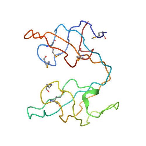
Crystal structure of the ydhK protein from Bacillus subtilis.
Vorobiev, S., Abashidze, M., Seetharaman, J., Mao, M., Xiao, R., Maglaqui, M., Owens, L.A., Cunningham, K., Zhao, L., Everett, J.K., Acton, T.B., Montelione, G.T., Tong, L., Hunt, J.F.To be published.