4EQM

Structural analysis of Staphylococcus aureus serine/threonine kinase PknB


Experimental Data Snapshot

  • Method: X-RAY DIFFRACTION
  • Resolution: 3.00 Å
  • R-Value Free: 0.246 
  • R-Value Work: 0.215 
  • R-Value Observed: 0.216 

wwPDB Validation   3D Report Full Report


Ligand Structure Quality Assessment 


This is version 1.2 of the entry. See complete history


Literature

Structural Analysis of Staphylococcus aureus Serine/Threonine Kinase PknB.

Rakette, S.Donat, S.Ohlsen, K.Stehle, T.

(2012) PLoS One 7: e39136-e39136

  • DOI: https://doi.org/10.1371/journal.pone.0039136
  • Primary Citation of Related Structures:  
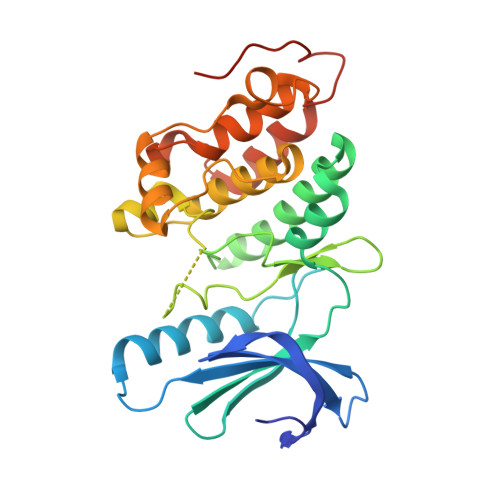
    4EQM

  • PubMed Abstract: 

    Effective treatment of infections caused by the bacterium Staphylococcus aureus remains a worldwide challenge, in part due to the constant emergence of new strains that are resistant to antibiotics. The serine/threonine kinase PknB is of particular relevance to the life cycle of S. aureus as it is involved in the regulation of purine biosynthesis, autolysis, and other central metabolic processes of the bacterium. We have determined the crystal structure of the kinase domain of PknB in complex with a non-hydrolyzable analog of the substrate ATP at 3.0 Å resolution. Although the purified PknB kinase is active in solution, it crystallized in an inactive, autoinhibited state. Comparison with other bacterial kinases provides insights into the determinants of catalysis, interactions of PknB with ligands, and the pathway of activation.


  • Organizational Affiliation

    Interfaculty Institute of Biochemistry, University of Tübingen, Tübingen, Germany.


Macromolecules
Find similar proteins by:  (by identity cutoff)  |  3D Structure
Entity ID: 1
MoleculeChains Sequence LengthOrganismDetailsImage
Protein kinase
A, B, C, D, E
A, B, C, D, E, F
294Staphylococcus aureus subsp. aureus N315Mutation(s): 0 
Gene Names: SA1063
UniProt
Find proteins for A0A0H3JME9 (Staphylococcus aureus (strain N315))
Explore A0A0H3JME9 
Go to UniProtKB:  A0A0H3JME9
Entity Groups  
Sequence Clusters30% Identity50% Identity70% Identity90% Identity95% Identity100% Identity
UniProt GroupA0A0H3JME9
Sequence Annotations
Expand
  • Reference Sequence
Small Molecules
Ligands 2 Unique
IDChains Name / Formula / InChI Key2D Diagram3D Interactions
ANP
Query on ANP

Download Ideal Coordinates CCD File 
G [auth A]
I [auth B]
K [auth C]
M [auth D]
N [auth E]
G [auth A],
I [auth B],
K [auth C],
M [auth D],
N [auth E],
O [auth F]
PHOSPHOAMINOPHOSPHONIC ACID-ADENYLATE ESTER
C10 H17 N6 O12 P3
PVKSNHVPLWYQGJ-KQYNXXCUSA-N
BEN
Query on BEN

Download Ideal Coordinates CCD File 
H [auth A],
J [auth B],
L [auth C]
BENZAMIDINE
C7 H8 N2
PXXJHWLDUBFPOL-UHFFFAOYSA-N
Experimental Data & Validation

Experimental Data

  • Method: X-RAY DIFFRACTION
  • Resolution: 3.00 Å
  • R-Value Free: 0.246 
  • R-Value Work: 0.215 
  • R-Value Observed: 0.216 
  • Space Group: C 1 2 1
Unit Cell:
Length ( Å )Angle ( ˚ )
a = 221.51α = 90
b = 127.55β = 89.96
c = 70.28γ = 90
Software Package:
Software NamePurpose
RemDAqdata collection
PHASERphasing
PHENIXrefinement
XDSdata reduction
XSCALEdata scaling

Structure Validation

View Full Validation Report


Ligand Structure Quality Assessment 


Entry History 

Deposition Data

Revision History  (Full details and data files)

  • Version 1.0: 2012-06-27
    Type: Initial release
  • Version 1.1: 2017-11-15
    Changes: Refinement description
  • Version 1.2: 2024-02-28
    Changes: Data collection, Database references, Derived calculations