4EOZ

Crystal structure of the SPOP BTB domain complexed with the Cul3 N-terminal domain


Experimental Data Snapshot

  • Method: X-RAY DIFFRACTION
  • Resolution: 2.40 Å
  • R-Value Free: 0.263 
  • R-Value Work: 0.211 
  • R-Value Observed: 0.214 

wwPDB Validation   3D Report Full Report


This is version 1.1 of the entry. See complete history


Literature

Adaptor protein self-assembly drives the control of a cullin-RING ubiquitin ligase.

Errington, W.J.Khan, M.Q.Bueler, S.A.Rubinstein, J.L.Chakrabartty, A.Prive, G.G.

(2012) Structure 20: 1141-1153

  • DOI: https://doi.org/10.1016/j.str.2012.04.009
  • Primary Citation of Related Structures:  
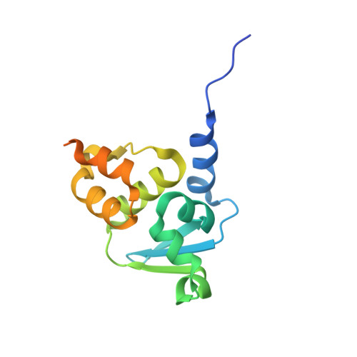
    4EOZ

  • PubMed Abstract: 

    The E3 ligases recruit substrate proteins for targeted ubiquitylation. Recent insights into the mechanisms of ubiquitylation demonstrate that E3 ligases can possess active regulatory properties beyond those of a simple assembly scaffold. Here, we describe the dimeric structure of the E3 ligase adaptor protein SPOP (speckle-type POZ protein) in complex with the N-terminal domain of Cul3 at 2.4 Å resolution. We find that SPOP forms large oligomers that can form heteromeric species with the closely related paralog SPOPL. In combination, SPOP and SPOPL (SPOP-like) form a molecular rheostat that can fine-tune E3 ubiquitin ligase activity by affecting the oligomeric state of the E3 complex. We propose that adaptor protein self-assembly provides a graded level of regulation of the SPOP/Cul3 E3 ligase toward its multiple protein substrates.


  • Organizational Affiliation

    Department of Biochemistry, University of Toronto, 1 Kings College Circle, Toronto, Ontario M5S 1A8, Canada.


Macromolecules
Find similar proteins by:  (by identity cutoff)  |  3D Structure
Entity ID: 1
MoleculeChains Sequence LengthOrganismDetailsImage
Speckle-type POZ proteinA,
B [auth C]
145Homo sapiensMutation(s): 0 
Gene Names: SPOP
UniProt & NIH Common Fund Data Resources
Find proteins for O43791 (Homo sapiens)
Explore O43791 
Go to UniProtKB:  O43791
PHAROS:  O43791
GTEx:  ENSG00000121067 
Entity Groups  
Sequence Clusters30% Identity50% Identity70% Identity90% Identity95% Identity100% Identity
UniProt GroupO43791
Sequence Annotations
Expand
  • Reference Sequence
Find similar proteins by:  (by identity cutoff)  |  3D Structure
Entity ID: 2
MoleculeChains Sequence LengthOrganismDetailsImage
Cullin-3C [auth B],
D
364Homo sapiensMutation(s): 2 
Gene Names: CUL3KIAA0617
UniProt & NIH Common Fund Data Resources
Find proteins for Q13618 (Homo sapiens)
Explore Q13618 
Go to UniProtKB:  Q13618
PHAROS:  Q13618
GTEx:  ENSG00000036257 
Entity Groups  
Sequence Clusters30% Identity50% Identity70% Identity90% Identity95% Identity100% Identity
UniProt GroupQ13618
Sequence Annotations
Expand
  • Reference Sequence
Small Molecules
Modified Residues  1 Unique
IDChains TypeFormula2D DiagramParent
MSE
Query on MSE
C [auth B],
D
L-PEPTIDE LINKINGC5 H11 N O2 SeMET
Experimental Data & Validation

Experimental Data

  • Method: X-RAY DIFFRACTION
  • Resolution: 2.40 Å
  • R-Value Free: 0.263 
  • R-Value Work: 0.211 
  • R-Value Observed: 0.214 
  • Space Group: C 1 2 1
Unit Cell:
Length ( Å )Angle ( ˚ )
a = 213.166α = 90
b = 76.944β = 108.34
c = 85.834γ = 90
Software Package:
Software NamePurpose
HKL-3000data collection
SOLVEphasing
PHENIXrefinement
HKL-3000data reduction
HKL-3000data scaling

Structure Validation

View Full Validation Report


Entry History 

Deposition Data

Revision History  (Full details and data files)

  • Version 1.0: 2012-05-30
    Type: Initial release
  • Version 1.1: 2013-01-09
    Changes: Database references