4EBV

Structure of Focal Adhesion Kinase catalytic domain in complex with novel allosteric inhibitor


Experimental Data Snapshot

  • Method: X-RAY DIFFRACTION
  • Resolution: 1.67 Å
  • R-Value Free: 0.236 
  • R-Value Work: 0.187 
  • R-Value Observed: 0.190 

wwPDB Validation   3D Report Full Report


Ligand Structure Quality Assessment 


This is version 1.4 of the entry. See complete history


Literature

Discovery and characterization of novel allosteric FAK inhibitors.

Iwatani, M.Iwata, H.Okabe, A.Skene, R.J.Tomita, N.Hayashi, Y.Aramaki, Y.Hosfield, D.J.Hori, A.Baba, A.Miki, H.

(2013) Eur J Med Chem 61: 49-60

  • DOI: https://doi.org/10.1016/j.ejmech.2012.06.035
  • Primary Citation of Related Structures:  
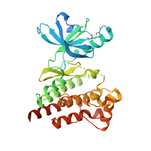
    4EBV, 4EBW

  • PubMed Abstract: 

    Focal adhesion kinase (FAK) regulates cell survival and proliferation pathways. Here we report the discovery of a highly selective series of 1,5-dihydropyrazolo[4,3-c][2,1]benzothiazines that demonstrate a novel mode of allosteric inhibition of FAK. These compounds showed slow dissociation from unphosphorylated FAK and were noncompetitive with ATP after long preincubation. Co-crystal structural analysis revealed that the compounds target a novel allosteric site within the C-lobe of the kinase domain, which induces disruption of ATP pocket formation leading to the inhibition of kinase activity. The potency of allosteric inhibition was reduced by phosphorylation of FAK. Coupled SAR analysis revealed that N-substitution of the fused pyrazole is critical to achieve allosteric binding and high selectivity among kinases.


  • Organizational Affiliation

    Pharmaceutical Research Division, Takeda Pharmaceutical Company, Ltd., 26-1, Muraoka-Higashi 2-chome, Fujisawa, Kanagawa 251-8555, Japan.


Macromolecules
Find similar proteins by:  (by identity cutoff)  |  3D Structure
Entity ID: 1
MoleculeChains Sequence LengthOrganismDetailsImage
Focal adhesion kinase 1304Homo sapiensMutation(s): 0 
Gene Names: PTK2FAKFAK1
EC: 2.7.10.2
UniProt & NIH Common Fund Data Resources
Find proteins for Q05397 (Homo sapiens)
Explore Q05397 
Go to UniProtKB:  Q05397
PHAROS:  Q05397
GTEx:  ENSG00000169398 
Entity Groups  
Sequence Clusters30% Identity50% Identity70% Identity90% Identity95% Identity100% Identity
UniProt GroupQ05397
Sequence Annotations
Expand
  • Reference Sequence
Small Molecules
Ligands 2 Unique
IDChains Name / Formula / InChI Key2D Diagram3D Interactions
0O7
Query on 0O7

Download Ideal Coordinates CCD File 
B [auth A]8-(4-ethylphenyl)-5-methyl-2,5-dihydropyrazolo[4,3-c][2,1]benzothiazine 4,4-dioxide
C18 H17 N3 O2 S
ISXAVIPPPMJTPN-UHFFFAOYSA-N
IPA
Query on IPA

Download Ideal Coordinates CCD File 
C [auth A],
D [auth A]
ISOPROPYL ALCOHOL
C3 H8 O
KFZMGEQAYNKOFK-UHFFFAOYSA-N
Binding Affinity Annotations 
IDSourceBinding Affinity
0O7 BindingDB:  4EBV IC50: min: 880, max: 3.00e+4 (nM) from 15 assay(s)
Binding MOAD:  4EBV IC50: 990 (nM) from 1 assay(s)
PDBBind:  4EBV IC50: 4200 (nM) from 1 assay(s)
Experimental Data & Validation

Experimental Data

  • Method: X-RAY DIFFRACTION
  • Resolution: 1.67 Å
  • R-Value Free: 0.236 
  • R-Value Work: 0.187 
  • R-Value Observed: 0.190 
  • Space Group: C 1 2 1
Unit Cell:
Length ( Å )Angle ( ˚ )
a = 74.222α = 90
b = 47.954β = 112.45
c = 83.281γ = 90
Software Package:
Software NamePurpose
HKL-2000data collection
MOLREPphasing
REFMACrefinement
HKL-2000data reduction
HKL-2000data scaling

Structure Validation

View Full Validation Report


Ligand Structure Quality Assessment 


Entry History 

Deposition Data

Revision History  (Full details and data files)

  • Version 1.0: 2012-08-22
    Type: Initial release
  • Version 1.1: 2013-01-02
    Changes: Database references
  • Version 1.2: 2013-01-16
    Changes: Database references
  • Version 1.3: 2013-03-27
    Changes: Database references
  • Version 1.4: 2023-09-13
    Changes: Data collection, Database references, Derived calculations, Refinement description