4DGG

c-SRC kinase domain in complex with RM-1-176


Experimental Data Snapshot

  • Method: X-RAY DIFFRACTION
  • Resolution: 2.65 Å
  • R-Value Free: 0.252 
  • R-Value Work: 0.208 
  • R-Value Observed: 0.210 

wwPDB Validation   3D Report Full Report


Ligand Structure Quality Assessment 


This is version 1.3 of the entry. See complete history


Literature

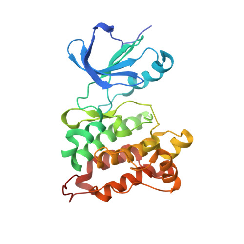
Active site profiling reveals coupling between domains in SRC-family kinases.

Krishnamurty, R.Brigham, J.L.Leonard, S.E.Ranjitkar, P.Larson, E.T.Dale, E.J.Merritt, E.A.Maly, D.J.

(2013) Nat Chem Biol 9: 43-50

  • DOI: https://doi.org/10.1038/nchembio.1118
  • Primary Citation of Related Structures:  
    4DGG

  • PubMed Abstract: 

    Protein kinases, key regulators of intracellular signal transduction, have emerged as an important class of drug targets. Chemical proteomic tools that facilitate the functional interrogation of protein kinase active sites are powerful reagents for studying the regulation of this large enzyme family and performing inhibitor selectivity screens. Here we describe a new crosslinking strategy that enables rapid and quantitative profiling of protein kinase active sites in lysates and live cells. Applying this methodology to the SRC-family kinases (SFKs) SRC and HCK led to the identification of a series of conformation-specific, ATP-competitive inhibitors that have a distinct preference for the autoinhibited forms of these kinases. Furthermore, we show that ligands that have this selectivity are able to modulate the ability of the regulatory domains of SRC and HCK to engage in intermolecular binding interactions. These studies provide insight into the regulation of this important family of tyrosine kinases.


  • Organizational Affiliation

    Department of Chemistry, University of Washington, Seattle, Washington, USA.


Macromolecules
Find similar proteins by:  (by identity cutoff)  |  3D Structure
Entity ID: 1
MoleculeChains Sequence LengthOrganismDetailsImage
Proto-oncogene tyrosine-protein kinase Src
A, B
286Gallus gallusMutation(s): 0 
Gene Names: SRC
EC: 2.7.10.2
UniProt
Find proteins for P00523 (Gallus gallus)
Explore P00523 
Go to UniProtKB:  P00523
Entity Groups  
Sequence Clusters30% Identity50% Identity70% Identity90% Identity95% Identity100% Identity
UniProt GroupP00523
Sequence Annotations
Expand
  • Reference Sequence
Small Molecules
Ligands 1 Unique
IDChains Name / Formula / InChI Key2D Diagram3D Interactions
I76
Query on I76

Download Ideal Coordinates CCD File 
C [auth A],
D [auth B]
3-{6-[(3-chlorobenzyl)oxy]naphthalen-2-yl}-1-(propan-2-yl)-1H-pyrazolo[3,4-d]pyrimidin-4-amine
C25 H22 Cl N5 O
MGARWHQGMCYTAL-UHFFFAOYSA-N
Binding Affinity Annotations 
IDSourceBinding Affinity
I76 PDBBind:  4DGG IC50: 800 (nM) from 1 assay(s)
BindingDB:  4DGG IC50: 280 (nM) from 1 assay(s)
Experimental Data & Validation

Experimental Data

  • Method: X-RAY DIFFRACTION
  • Resolution: 2.65 Å
  • R-Value Free: 0.252 
  • R-Value Work: 0.208 
  • R-Value Observed: 0.210 
  • Space Group: P 1
Unit Cell:
Length ( Å )Angle ( ˚ )
a = 42.17α = 78.73
b = 63.48β = 88.48
c = 74γ = 89.93
Software Package:
Software NamePurpose
SCALAdata scaling
REFMACrefinement
PDB_EXTRACTdata extraction
Blu-Icedata collection
MOSFLMdata reduction

Structure Validation

View Full Validation Report


Ligand Structure Quality Assessment 


Entry History 

Deposition Data

Revision History  (Full details and data files)

  • Version 1.0: 2012-10-10
    Type: Initial release
  • Version 1.1: 2012-11-21
    Changes: Database references
  • Version 1.2: 2013-01-02
    Changes: Database references
  • Version 1.3: 2024-02-28
    Changes: Data collection, Database references, Derived calculations, Refinement description