4APF

Crystal structure of the human KLHL11-Cul3 complex at 3.1A resolution


Experimental Data Snapshot

  • Method: X-RAY DIFFRACTION
  • Resolution: 3.10 Å
  • R-Value Free: 0.222 
  • R-Value Work: 0.193 
  • R-Value Observed: 0.195 

wwPDB Validation   3D Report Full Report


This is version 1.3 of the entry. See complete history


Literature

Structural Basis for Cul3 Assembly with the Btb-Kelch Family of E3 Ubiquitin Ligases.

Canning, P.Cooper, C.D.O.Krojer, T.Murray, J.W.Pike, A.C.W.Chaikuad, A.Keates, T.Thangaratnarajah, C.Hojzan, V.Marsden, B.D.Gileadi, O.Knapp, S.von Delft, F.Bullock, A.N.

(2013) J Biol Chem 288: 7803

  • DOI: https://doi.org/10.1074/jbc.M112.437996
  • Primary Citation of Related Structures:  
    2VPJ, 2XN4, 3II7, 4AP2, 4APF, 4ASC

  • PubMed Abstract: 

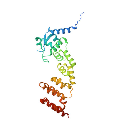
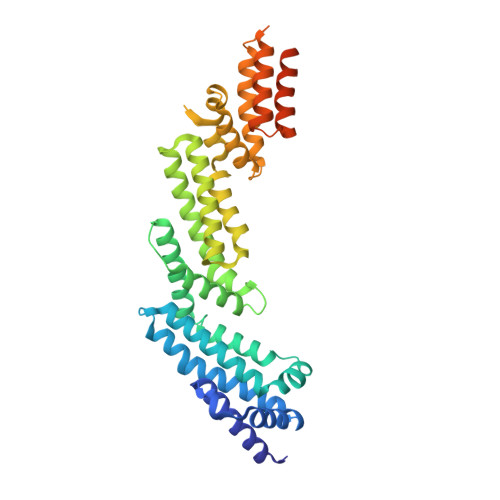
    Cullin-RING ligases are multisubunit E3 ubiquitin ligases that recruit substrate-specific adaptors to catalyze protein ubiquitylation. Cul3-based Cullin-RING ligases are uniquely associated with BTB adaptors that incorporate homodimerization, Cul3 assembly, and substrate recognition into a single multidomain protein, of which the best known are BTB-BACK-Kelch domain proteins, including KEAP1. Cul3 assembly requires a BTB protein "3-box" motif, analogous to the F-box and SOCS box motifs of other Cullin-based E3s. To define the molecular basis for this assembly and the overall architecture of the E3, we determined the crystal structures of the BTB-BACK domains of KLHL11 both alone and in complex with Cul3, along with the Kelch domain structures of KLHL2 (Mayven), KLHL7, KLHL12, and KBTBD5. We show that Cul3 interaction is dependent on a unique N-terminal extension sequence that packs against the 3-box in a hydrophobic groove centrally located between the BTB and BACK domains. Deletion of this N-terminal region results in a 30-fold loss in affinity. The presented data offer a model for the quaternary assembly of this E3 class that supports the bivalent capture of Nrf2 and reveals potential new sites for E3 inhibitor design.


  • Organizational Affiliation

    Structural Genomics Consortium, University of Oxford, Oxford OX3 7DQ, United Kingdom.


Macromolecules
Find similar proteins by:  (by identity cutoff)  |  3D Structure
Entity ID: 1
MoleculeChains Sequence LengthOrganismDetailsImage
KELCH-LIKE PROTEIN 11297Homo sapiensMutation(s): 0 
UniProt & NIH Common Fund Data Resources
Find proteins for Q9NVR0 (Homo sapiens)
Explore Q9NVR0 
Go to UniProtKB:  Q9NVR0
PHAROS:  Q9NVR0
GTEx:  ENSG00000178502 
Entity Groups  
Sequence Clusters30% Identity50% Identity70% Identity90% Identity95% Identity100% Identity
UniProt GroupQ9NVR0
Sequence Annotations
Expand
  • Reference Sequence
Find similar proteins by:  (by identity cutoff)  |  3D Structure
Entity ID: 2
MoleculeChains Sequence LengthOrganismDetailsImage
CULLIN 3388Homo sapiensMutation(s): 2 
UniProt & NIH Common Fund Data Resources
Find proteins for Q13618 (Homo sapiens)
Explore Q13618 
Go to UniProtKB:  Q13618
PHAROS:  Q13618
GTEx:  ENSG00000036257 
Entity Groups  
Sequence Clusters30% Identity50% Identity70% Identity90% Identity95% Identity100% Identity
UniProt GroupQ13618
Sequence Annotations
Expand
  • Reference Sequence
Experimental Data & Validation

Experimental Data

  • Method: X-RAY DIFFRACTION
  • Resolution: 3.10 Å
  • R-Value Free: 0.222 
  • R-Value Work: 0.193 
  • R-Value Observed: 0.195 
  • Space Group: C 1 2 1
Unit Cell:
Length ( Å )Angle ( ˚ )
a = 238.64α = 90
b = 41.44β = 110.2
c = 147.78γ = 90
Software Package:
Software NamePurpose
BUSTERrefinement
MOSFLMdata reduction
SCALAdata scaling
SHARPphasing

Structure Validation

View Full Validation Report


Entry History 

Revision History  (Full details and data files)

  • Version 1.0: 2012-05-09
    Type: Initial release
  • Version 1.1: 2013-02-06
    Changes: Database references, Refinement description, Structure summary
  • Version 1.2: 2013-03-27
    Changes: Database references
  • Version 1.3: 2018-01-24
    Changes: Database references, Structure summary