3ZC6

Crystal structure of JAK3 kinase domain in complex with an indazole substituted pyrrolopyrazine inhibitor


Experimental Data Snapshot

  • Method: X-RAY DIFFRACTION
  • Resolution: 2.42 Å
  • R-Value Free: 0.254 
  • R-Value Work: 0.191 
  • R-Value Observed: 0.193 

wwPDB Validation   3D Report Full Report


Ligand Structure Quality Assessment 


This is version 1.0 of the entry. See complete history


Literature

Strategic Use of Conformational Bias and Structure Based Design to Identify Potent Jak3 Inhibitors with Improved Selectivity Against the Jak Family and the Kinome.

Lynch, S.M.Devicente, J.Hermann, J.C.Jaime-Figueroa, S.Jin, S.Kuglstatter, A.Li, H.Lovey, A.Menke, J.Niu, L.Patel, V.Roy, D.Soth, M.Steiner, S.Tivitmahaisoon, P.Vu, M.D.Yee, C.

(2013) Bioorg Med Chem Lett 23: 2793

  • DOI: https://doi.org/10.1016/j.bmcl.2013.02.012
  • Primary Citation of Related Structures:  
    3ZC6

  • PubMed Abstract: 

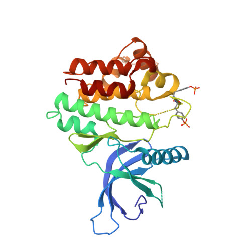
    Using a structure based design approach we have identified a series of indazole substituted pyrrolopyrazines, which are potent inhibitors of JAK3. Intramolecular electronic repulsion was used as a strategy to induce a strong conformational bias within the ligand. Compounds bearing this conformation participated in a favorable hydrophobic interaction with a cysteine residue in the JAK3 binding pocket, which imparted high selectivity versus the kinome and improved selectivity within the JAK family.


  • Organizational Affiliation

    Discovery Chemistry, Hoffmann-La Roche, pRED, Pharma Research & Early Development, 340 Kingsland Street, Nutley, NJ 07110, USA. stephen.lynch@roche.com


Macromolecules
Find similar proteins by:  (by identity cutoff)  |  3D Structure
Entity ID: 1
MoleculeChains Sequence LengthOrganismDetailsImage
TYROSINE-PROTEIN KINASE JAK3
A, B, C, D
288Homo sapiensMutation(s): 2 
EC: 2.7.10.2 (PDB Primary Data), 2.7.1.112 (PDB Primary Data)
UniProt & NIH Common Fund Data Resources
Find proteins for P52333 (Homo sapiens)
Explore P52333 
Go to UniProtKB:  P52333
PHAROS:  P52333
GTEx:  ENSG00000105639 
Entity Groups  
Sequence Clusters30% Identity50% Identity70% Identity90% Identity95% Identity100% Identity
UniProt GroupP52333
Sequence Annotations
Expand
  • Reference Sequence
Small Molecules
Ligands 4 Unique
IDChains Name / Formula / InChI Key2D Diagram3D Interactions
VFC
Query on VFC

Download Ideal Coordinates CCD File 
E [auth A],
J [auth B],
M [auth C],
O [auth D]
N-[(2R)-1-(3-cyanoazetidin-1-yl)-1-oxidanylidene-propan-2-yl]-2-(6-fluoranyl-1-methyl-indazol-3-yl)-5H-pyrrolo[2,3-b]pyrazine-7-carboxamide
C22 H19 F N8 O2
AGTYRXOJPORGGM-LLVKDONJSA-N
PG4
Query on PG4

Download Ideal Coordinates CCD File 
K [auth B],
N [auth C]
TETRAETHYLENE GLYCOL
C8 H18 O5
UWHCKJMYHZGTIT-UHFFFAOYSA-N
PEG
Query on PEG

Download Ideal Coordinates CCD File 
F [auth A],
L [auth B]
DI(HYDROXYETHYL)ETHER
C4 H10 O3
MTHSVFCYNBDYFN-UHFFFAOYSA-N
CL
Query on CL

Download Ideal Coordinates CCD File 
G [auth A],
H [auth A],
I [auth A],
P [auth D]
CHLORIDE ION
Cl
VEXZGXHMUGYJMC-UHFFFAOYSA-M
Modified Residues  1 Unique
IDChains TypeFormula2D DiagramParent
PTR
Query on PTR
A, B, C, D
L-PEPTIDE LINKINGC9 H12 N O6 PTYR
Binding Affinity Annotations 
IDSourceBinding Affinity
VFC BindingDB:  3ZC6 IC50: min: 0.6, max: 150 (nM) from 2 assay(s)
Binding MOAD:  3ZC6 IC50: 0.6 (nM) from 1 assay(s)
PDBBind:  3ZC6 IC50: 0.6 (nM) from 1 assay(s)
Experimental Data & Validation

Experimental Data

  • Method: X-RAY DIFFRACTION
  • Resolution: 2.42 Å
  • R-Value Free: 0.254 
  • R-Value Work: 0.191 
  • R-Value Observed: 0.193 
  • Space Group: P 1 21 1
Unit Cell:
Length ( Å )Angle ( ˚ )
a = 57.17α = 90
b = 117.725β = 97.66
c = 105.831γ = 90
Software Package:
Software NamePurpose
REFMACrefinement
XDSdata reduction
XSCALEdata scaling
REFMACphasing

Structure Validation

View Full Validation Report


Ligand Structure Quality Assessment 


Entry History 

Deposition Data

Revision History  (Full details and data files)

  • Version 1.0: 2013-09-25
    Type: Initial release