3VF8

Crystal Structure of Spleen Tyrosine Kinase Syk Catalytic Domain with Pyrazolylbenzimidazole Inhibitor 416


Experimental Data Snapshot

  • Method: X-RAY DIFFRACTION
  • Resolution: 2.08 Å
  • R-Value Free: 0.241 
  • R-Value Work: 0.215 
  • R-Value Observed: 0.217 

wwPDB Validation   3D Report Full Report


Ligand Structure Quality Assessment 


This is version 1.2 of the entry. See complete history


Literature

X-ray crystallographic structure-based design of selective thienopyrazole inhibitors for interleukin-2-inducible tyrosine kinase.

McLean, L.R.Zhang, Y.Zaidi, N.Bi, X.Wang, R.Dharanipragada, R.Jurcak, J.G.Gillespy, T.A.Zhao, Z.Musick, K.Y.Choi, Y.M.Barrague, M.Peppard, J.Smicker, M.Duguid, M.Parkar, A.Fordham, J.Kominos, D.

(2012) Bioorg Med Chem Lett 22: 3296-3300

  • DOI: https://doi.org/10.1016/j.bmcl.2012.03.016
  • Primary Citation of Related Structures:  
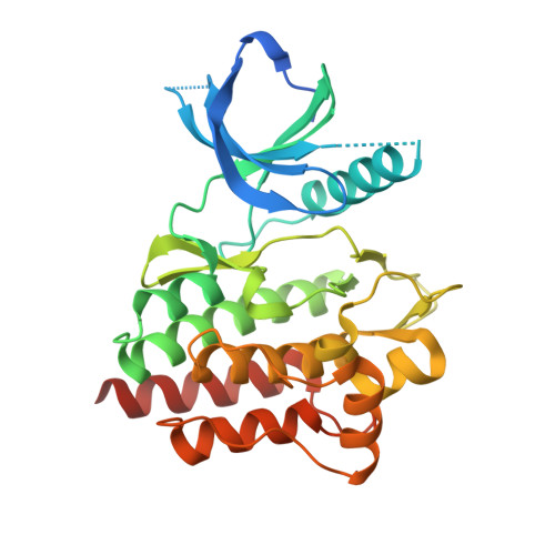
    3V5J, 3V5L, 3V8T, 3V8W, 3VF8, 3VF9

  • PubMed Abstract: 

    Beginning with a screening hit, unique thienopyrazole-indole inhibitors of Itk (interleukin-2-inducible tyrosine kinase) were designed, synthesized, and crystallized in the target kinase. Although initial compounds were highly active in Itk, they were not selective. Increasing the steric bulk around a tertiary alcohol at the 5-indole position dramatically improved selectivity toward Lyk and Syk, but not Txk. Substitutions at the 3- and 4-indole positions gave less active compounds that remained poorly selective. A difluoromethyl substitution at the 5-position of the thienopyrazole led to a highly potent and selective compound. Phenyl at this position reduced activity and selectivity while pushing the side-chains of Lys-391 and Asp-500 away from the binding pocket. Novel and selective thienopyrazole inhibitors of Itk were designed as a result of combining structure-based design and medicinal chemistry.


  • Organizational Affiliation

    Molecular Innovative Therapeutics, Sanofi US, 1041 Route 202/206 N, Bridgewater, NJ 08807, United States. Larry.McLean@sanofi.com


Macromolecules
Find similar proteins by:  (by identity cutoff)  |  3D Structure
Entity ID: 1
MoleculeChains Sequence LengthOrganismDetailsImage
Tyrosine-protein kinase SYK299Homo sapiensMutation(s): 0 
Gene Names: SYK
EC: 2.7.10.2
UniProt & NIH Common Fund Data Resources
Find proteins for P43405 (Homo sapiens)
Explore P43405 
Go to UniProtKB:  P43405
PHAROS:  P43405
GTEx:  ENSG00000165025 
Entity Groups  
Sequence Clusters30% Identity50% Identity70% Identity90% Identity95% Identity100% Identity
UniProt GroupP43405
Sequence Annotations
Expand
  • Reference Sequence
Small Molecules
Ligands 1 Unique
IDChains Name / Formula / InChI Key2D Diagram3D Interactions
0JE
Query on 0JE

Download Ideal Coordinates CCD File 
B [auth A]3-[5-(5-ethoxy-6-fluoro-1H-benzimidazol-2-yl)-1H-pyrazol-4-yl]-1,1-diethylurea
C17 H21 F N6 O2
UJXIKUVGGQECLJ-UHFFFAOYSA-N
Binding Affinity Annotations 
IDSourceBinding Affinity
0JE Binding MOAD:  3VF8 IC50: 150 (nM) from 1 assay(s)
PDBBind:  3VF8 IC50: 150 (nM) from 1 assay(s)
Experimental Data & Validation

Experimental Data

  • Method: X-RAY DIFFRACTION
  • Resolution: 2.08 Å
  • R-Value Free: 0.241 
  • R-Value Work: 0.215 
  • R-Value Observed: 0.217 
  • Space Group: P 21 21 21
Unit Cell:
Length ( Å )Angle ( ˚ )
a = 39.743α = 90
b = 84.952β = 90
c = 89.734γ = 90
Software Package:
Software NamePurpose
SCALEPACKdata scaling
PHASERphasing
BUSTER-TNTrefinement
PDB_EXTRACTdata extraction
HKL-2000data collection
DENZOdata reduction
BUSTERrefinement

Structure Validation

View Full Validation Report


Ligand Structure Quality Assessment 


Entry History 

Deposition Data

Revision History  (Full details and data files)

  • Version 1.0: 2012-05-02
    Type: Initial release
  • Version 1.1: 2017-11-15
    Changes: Refinement description
  • Version 1.2: 2024-02-28
    Changes: Data collection, Database references, Derived calculations