3V3V

Structural and functional analysis of quercetagetin, a natural JNK1 inhibitor


Experimental Data Snapshot

  • Method: X-RAY DIFFRACTION
  • Resolution: 2.70 Å
  • R-Value Free: 0.266 
  • R-Value Work: 0.227 
  • R-Value Observed: 0.228 

wwPDB Validation   3D Report Full Report


Ligand Structure Quality Assessment 


This is version 1.2 of the entry. See complete history


Literature

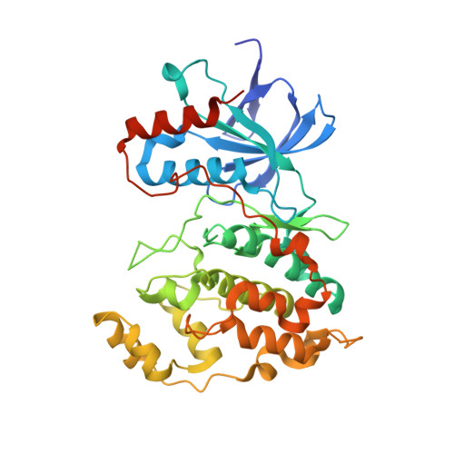
Structural and Functional Analysis of the Natural JNK1 Inhibitor Quercetagetin.

Baek, S.Kang, N.J.Popowicz, G.M.Arciniega, M.Jung, S.K.Byun, S.Song, N.R.Heo, Y.S.Kim, B.Y.Lee, H.J.Holak, T.A.Augustin, M.Bode, A.M.Huber, R.Dong, Z.Lee, K.W.

(2013) J Mol Biol 425: 411-423

  • DOI: https://doi.org/10.1016/j.jmb.2012.10.019
  • Primary Citation of Related Structures:  
    3V3V

  • PubMed Abstract: 

    c-Jun NH2-terminal kinases (JNKs) and phosphatidylinositol 3-kinase (PI3-K) play critical roles in chronic diseases such as cancer, type II diabetes, and obesity. We describe here the binding of quercetagetin (3,3',4',5,6,7-hydroxyflavone), related flavonoids, and SP600125 to JNK1 and PI3-K by ATP-competitive and immobilized metal ion affinity-based fluorescence polarization assays and measure the effect of quercetagetin on JNK1 and PI3-K activities. Quercetagetin attenuated the phosphorylation of c-Jun and AKT, suppressed AP-1 and NF-κB promoter activities, and also reduced cell transformation. It attenuated tumor incidence and reduced tumor volumes in a two-stage skin carcinogenesis mouse model. Our crystallographic structure determination data show that quercetagetin binds to the ATP-binding site of JNK1. Notably, the interaction between Lys55, Asp169, and Glu73 of JNK1 and the catechol moiety of quercetagetin reorients the N-terminal lobe of JNK1, thereby improving compatibility of the ligand with its binding site. The results of a theoretical docking study suggest a binding mode of PI3-K with the hydroxyl groups of the catechol moiety forming hydrogen bonds with the side chains of Asp964 and Asp841 in the p110γ catalytic subunit. These interactions could contribute to the high inhibitory activity of quercetagetin against PI3-K. Our study suggests the potential use of quercetagetin in the prevention or therapy of cancer and other chronic diseases.


  • Organizational Affiliation

    Max Planck Institute for Biochemistry, Am Klopferspitz 18, 82152 Martinsried, Germany.


Macromolecules
Find similar proteins by:  (by identity cutoff)  |  3D Structure
Entity ID: 1
MoleculeChains Sequence LengthOrganismDetailsImage
Mitogen-activated protein kinase 8379Homo sapiensMutation(s): 0 
Gene Names: MAPK8JNK1PRKM8SAPK1SAPK1C
EC: 2.7.11.24
UniProt & NIH Common Fund Data Resources
Find proteins for P45983 (Homo sapiens)
Explore P45983 
Go to UniProtKB:  P45983
PHAROS:  P45983
GTEx:  ENSG00000107643 
Entity Groups  
Sequence Clusters30% Identity50% Identity70% Identity90% Identity95% Identity100% Identity
UniProt GroupP45983
Sequence Annotations
Expand
  • Reference Sequence

Find similar proteins by:  Sequence   |   3D Structure  

Entity ID: 2
MoleculeChains Sequence LengthOrganismDetailsImage
C-Jun-amino-terminal kinase-interacting protein 111Mus musculusMutation(s): 0 
UniProt
Find proteins for Q9WVI9 (Mus musculus)
Explore Q9WVI9 
Go to UniProtKB:  Q9WVI9
Entity Groups  
Sequence Clusters30% Identity50% Identity70% Identity90% Identity95% Identity100% Identity
UniProt GroupQ9WVI9
Sequence Annotations
Expand
  • Reference Sequence
Small Molecules
Ligands 3 Unique
IDChains Name / Formula / InChI Key2D Diagram3D Interactions
MYU
Query on MYU

Download Ideal Coordinates CCD File 
C [auth A]3,5,6,7-TETRAHYDROXY-2-(3,4-DIHYDROXYPHENYL)-4H-CHROMEN-4-ONE
C15 H10 O8
ZVOLCUVKHLEPEV-UHFFFAOYSA-N
SO4
Query on SO4

Download Ideal Coordinates CCD File 
D [auth A]
E [auth A]
F [auth A]
G [auth A]
H [auth A]
D [auth A],
E [auth A],
F [auth A],
G [auth A],
H [auth A],
I [auth A],
K [auth B]
SULFATE ION
O4 S
QAOWNCQODCNURD-UHFFFAOYSA-L
CL
Query on CL

Download Ideal Coordinates CCD File 
J [auth A]CHLORIDE ION
Cl
VEXZGXHMUGYJMC-UHFFFAOYSA-M
Binding Affinity Annotations 
IDSourceBinding Affinity
MYU PDBBind:  3V3V IC50: 4600 (nM) from 1 assay(s)
Experimental Data & Validation

Experimental Data

  • Method: X-RAY DIFFRACTION
  • Resolution: 2.70 Å
  • R-Value Free: 0.266 
  • R-Value Work: 0.227 
  • R-Value Observed: 0.228 
  • Space Group: I 4 2 2
Unit Cell:
Length ( Å )Angle ( ˚ )
a = 172.37α = 90
b = 172.37β = 90
c = 86.04γ = 90
Software Package:
Software NamePurpose
MAR345dtbdata collection
MOLREPphasing
REFMACrefinement
XDSdata reduction
XSCALEdata scaling

Structure Validation

View Full Validation Report


Ligand Structure Quality Assessment 


Entry History 

Revision History  (Full details and data files)

  • Version 1.0: 2012-12-05
    Type: Initial release
  • Version 1.1: 2013-01-30
    Changes: Database references
  • Version 1.2: 2023-09-13
    Changes: Data collection, Database references, Derived calculations, Refinement description