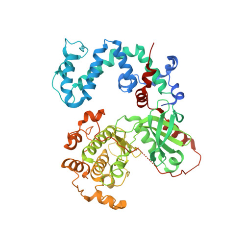
3T8O

Rhodopsin kinase (GRK1) L166K mutant at 2.5A resolution


Experimental Data Snapshot

  • Method: X-RAY DIFFRACTION
  • Resolution: 2.50 Å
  • R-Value Free: 0.245 
  • R-Value Work: 0.207 
  • R-Value Observed: 0.209 

wwPDB Validation   3D Report Full Report


Ligand Structure Quality Assessment 


This is version 1.3 of the entry. See complete history


Literature

Structure of a monomeric variant of rhodopsin kinase at 2.5 A resolution.

Tesmer, J.J.Nance, M.R.Singh, P.Lee, H.

(2012) Acta Crystallogr Sect F Struct Biol Cryst Commun 68: 622-625

  • DOI: https://doi.org/10.1107/S1744309112017435
  • Primary Citation of Related Structures:  
    3T8O

  • PubMed Abstract: 

    G protein-coupled receptor kinase 1 (GRK1 or rhodopsin kinase) phosphorylates activated rhodopsin and initiates a cascade of events that results in the termination of phototransduction by the receptor. Although GRK1 seems to be a monomer in solution, seven prior crystal structures of GRK1 revealed a similar domain-swapped dimer interface involving the C-terminus of the enzyme. The influence of this interface on the overall conformation of GRK1 is not known. To address this question, the crystalline dimer interface was disrupted with a L166K mutation and the structure of GRK1-L166K was determined in complex with Mg(2+) · ATP to 2.5 Å resolution. GRK1-L166K crystallized in a novel space group as a monomer and exhibited little overall conformational difference from prior structures of GRK1, although the C-terminal domain-swapped region had reorganized owing to loss of the dimer interface.


  • Organizational Affiliation

    Life Sciences Institute and the Department of Pharmacology, University of Michigan, 210 Washtenaw Avenue, Ann Arbor, MI 48109-2216, USA. tesmerjj@umich.edu


Macromolecules
Find similar proteins by:  (by identity cutoff)  |  3D Structure
Entity ID: 1
MoleculeChains Sequence LengthOrganismDetailsImage
Rhodopsin kinase543Bos taurusMutation(s): 1 
Gene Names: GRK1RHOK
EC: 2.7.11.14
UniProt
Find proteins for P28327 (Bos taurus)
Explore P28327 
Go to UniProtKB:  P28327
Entity Groups  
Sequence Clusters30% Identity50% Identity70% Identity90% Identity95% Identity100% Identity
UniProt GroupP28327
Sequence Annotations
Expand
  • Reference Sequence
Experimental Data & Validation

Experimental Data

  • Method: X-RAY DIFFRACTION
  • Resolution: 2.50 Å
  • R-Value Free: 0.245 
  • R-Value Work: 0.207 
  • R-Value Observed: 0.209 
  • Space Group: I 2 2 2
Unit Cell:
Length ( Å )Angle ( ˚ )
a = 55.541α = 90
b = 149.808β = 90
c = 190.879γ = 90
Software Package:
Software NamePurpose
DENZOdata reduction
SCALEPACKdata scaling
REFMACrefinement
PDB_EXTRACTdata extraction

Structure Validation

View Full Validation Report


Ligand Structure Quality Assessment 


Entry History 

Deposition Data

Revision History  (Full details and data files)

  • Version 1.0: 2012-06-06
    Type: Initial release
  • Version 1.1: 2013-01-23
    Changes: Database references
  • Version 1.2: 2017-11-08
    Changes: Refinement description
  • Version 1.3: 2024-02-28
    Changes: Data collection, Database references, Derived calculations