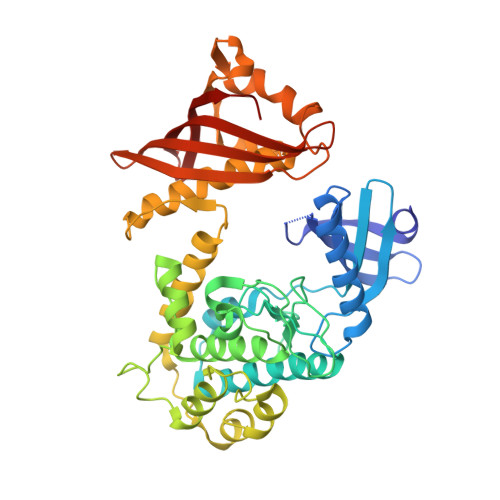
3SOA

Full-length human CaMKII


Experimental Data Snapshot

  • Method: X-RAY DIFFRACTION
  • Resolution: 3.55 Å
  • R-Value Free: 0.327 
  • R-Value Work: 0.273 
  • R-Value Observed: 0.279 

wwPDB Validation   3D Report Full Report


Ligand Structure Quality Assessment 


This is version 1.3 of the entry. See complete history


Literature

A Mechanism for Tunable Autoinhibition in the Structure of a Human Ca(2+)/Calmodulin- Dependent Kinase II Holoenzyme.

Chao, L.H.Stratton, M.M.Lee, I.H.Rosenberg, O.S.Levitz, J.Mandell, D.J.Kortemme, T.Groves, J.T.Schulman, H.Kuriyan, J.

(2011) Cell 146: 732-745

  • DOI: https://doi.org/10.1016/j.cell.2011.07.038
  • Primary Citation of Related Structures:  
    3SOA

  • PubMed Abstract: 

    Calcium/calmodulin-dependent kinase II (CaMKII) forms a highly conserved dodecameric assembly that is sensitive to the frequency of calcium pulse trains. Neither the structure of the dodecameric assembly nor how it regulates CaMKII are known. We present the crystal structure of an autoinhibited full-length human CaMKII holoenzyme, revealing an unexpected compact arrangement of kinase domains docked against a central hub, with the calmodulin-binding sites completely inaccessible. We show that this compact docking is important for the autoinhibition of the kinase domains and for setting the calcium response of the holoenzyme. Comparison of CaMKII isoforms, which differ in the length of the linker between the kinase domain and the hub, demonstrates that these interactions can be strengthened or weakened by changes in linker length. This equilibrium between autoinhibited states provides a simple mechanism for tuning the calcium response without changes in either the hub or the kinase domains.


  • Organizational Affiliation

    Department of Molecular and Cell Biology, University of California, Berkeley, CA 94720, USA.


Macromolecules
Find similar proteins by:  (by identity cutoff)  |  3D Structure
Entity ID: 1
MoleculeChains Sequence LengthOrganismDetailsImage
Calcium/calmodulin-dependent protein kinase type II subunit alpha with a beta 7 linker444Homo sapiensMutation(s): 3 
Gene Names: camk2acamkakiaa0968camk2bcamkb
EC: 2.7.11.17
UniProt & NIH Common Fund Data Resources
Find proteins for Q9UQM7 (Homo sapiens)
Explore Q9UQM7 
Go to UniProtKB:  Q9UQM7
PHAROS:  Q9UQM7
GTEx:  ENSG00000070808 
Entity Groups  
Sequence Clusters30% Identity50% Identity70% Identity90% Identity95% Identity100% Identity
UniProt GroupQ9UQM7
Sequence Annotations
Expand
  • Reference Sequence
Small Molecules
Ligands 1 Unique
IDChains Name / Formula / InChI Key2D Diagram3D Interactions
DB8
Query on DB8

Download Ideal Coordinates CCD File 
B [auth A]4-[(2,4-dichloro-5-methoxyphenyl)amino]-6-methoxy-7-[3-(4-methylpiperazin-1-yl)propoxy]quinoline-3-carbonitrile
C26 H29 Cl2 N5 O3
UBPYILGKFZZVDX-UHFFFAOYSA-N
Binding Affinity Annotations 
IDSourceBinding Affinity
DB8 BindingDB:  3SOA Kd: 4500 (nM) from 1 assay(s)
Experimental Data & Validation

Experimental Data

  • Method: X-RAY DIFFRACTION
  • Resolution: 3.55 Å
  • R-Value Free: 0.327 
  • R-Value Work: 0.273 
  • R-Value Observed: 0.279 
  • Space Group: P 6 2 2
Unit Cell:
Length ( Å )Angle ( ˚ )
a = 155.72α = 90
b = 155.72β = 90
c = 106.16γ = 120
Software Package:
Software NamePurpose
HKL-2000data collection
PHASERphasing
PHENIXrefinement
DENZOdata reduction
SCALAdata scaling

Structure Validation

View Full Validation Report


Ligand Structure Quality Assessment 


Entry History 

Deposition Data

Revision History  (Full details and data files)

  • Version 1.0: 2011-08-31
    Type: Initial release
  • Version 1.1: 2011-09-07
    Changes: Non-polymer description, Structure summary
  • Version 1.2: 2011-09-14
    Changes: Database references, Structure summary
  • Version 1.3: 2023-09-13
    Changes: Data collection, Database references, Derived calculations, Refinement description