3SHR

Crystal Structure of cGMP-dependent Protein Kinase Reveals Novel Site of Interchain Communication


Experimental Data Snapshot

  • Method: X-RAY DIFFRACTION
  • Resolution: 2.50 Å
  • R-Value Free: 0.277 
  • R-Value Work: 0.221 
  • R-Value Observed: 0.221 

wwPDB Validation   3D Report Full Report


Ligand Structure Quality Assessment 


This is version 1.1 of the entry. See complete history


Literature

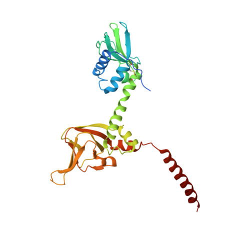
Crystal Structure of cGMP-Dependent Protein Kinase Reveals Novel Site of Interchain Communication.

Osborne, B.W.Wu, J.McFarland, C.J.Nickl, C.K.Sankaran, B.Casteel, D.E.Woods, V.L.Kornev, A.P.Taylor, S.S.Dostmann, W.R.

(2011) Structure 19: 1317-1327

  • DOI: https://doi.org/10.1016/j.str.2011.06.012
  • Primary Citation of Related Structures:  
    3SHR

  • PubMed Abstract: 

    The cGMP-dependent protein kinase (PKG) serves as an integral component of second messenger signaling in a number of biological contexts including cell differentiation, memory, and vasodilation. PKG is homodimeric and large conformational changes accompany cGMP binding. However, the structure of PKG and the molecular mechanisms associated with protomer communication following cGMP-induced activation remain unknown. Here, we report the 2.5 Å crystal structure of a regulatory domain construct (aa 78-355) containing both cGMP binding sites of PKG Iα. A distinct and segregated architecture with an extended central helix separates the two cGMP binding domains. Additionally, a previously uncharacterized helical domain (switch helix) promotes the formation of a hydrophobic interface between protomers. Mutational disruption of this interaction in full-length PKG implicates the switch helix as a critical site of dimer communication in PKG biology. These results offer new structural insight into the mechanism of allosteric PKG activation.


  • Organizational Affiliation

    Department of Pharmacology, College of Medicine, University of Vermont, Burlington, VT 05405, USA.


Macromolecules
Find similar proteins by:  (by identity cutoff)  |  3D Structure
Entity ID: 1
MoleculeChains Sequence LengthOrganismDetailsImage
cGMP-dependent protein kinase 1
A, B
299Bos taurusMutation(s): 0 
Gene Names: PRKG1PRKG1BPRKGR1APRKGR1B
EC: 2.7.11.12
UniProt
Find proteins for P00516 (Bos taurus)
Explore P00516 
Go to UniProtKB:  P00516
Entity Groups  
Sequence Clusters30% Identity50% Identity70% Identity90% Identity95% Identity100% Identity
UniProt GroupP00516
Sequence Annotations
Expand
  • Reference Sequence
Experimental Data & Validation

Experimental Data

  • Method: X-RAY DIFFRACTION
  • Resolution: 2.50 Å
  • R-Value Free: 0.277 
  • R-Value Work: 0.221 
  • R-Value Observed: 0.221 
  • Space Group: C 1 2 1
Unit Cell:
Length ( Å )Angle ( ˚ )
a = 180.156α = 90
b = 66.039β = 113.84
c = 81.55γ = 90
Software Package:
Software NamePurpose
HKL-2000data collection
CNSrefinement
HKL-2000data reduction

Structure Validation

View Full Validation Report


Ligand Structure Quality Assessment 


Entry History 

Deposition Data

Revision History  (Full details and data files)

  • Version 1.0: 2011-09-21
    Type: Initial release
  • Version 1.1: 2023-09-13
    Changes: Data collection, Database references, Derived calculations, Refinement description