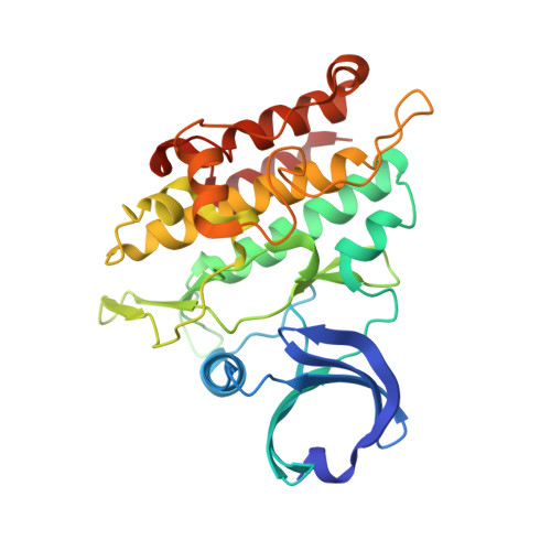
Crystal structure of the ACVR1 kinase domain in complex with the imidazo[1,2-b]pyridazine inhibitor K00507
Chaikuad, A., Sanvitale, C., Cooper, C., Mahajan, P., Daga, N., Petrie, K., Alfano, I., Gileadi, O., Fedorov, O., Allerston, C., Krojer, T., von Delft, F., Weigelt, J., Arrowsmith, C.H., Edwards, A.M., Bountra, C., Bullock, A.To be published.