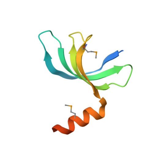
X-ray crystal structure of protein SP_0782 (7-79) from Streptococcus pneumoniae. Northeast Structural Genomics Consortium Target SpR104
Kuzin, A., Abashidze, M., Lew, S., Seetharaman, J., Patel, P., Xiao, R., Ciccosanti, C., Lee, D., Everett, J.K., Nair, R., Acton, T.B., Rost, B., Montelione, G.T., Tong, L., Hunt, J.F.To be published.