3N5U

Crystal structure of an Rb C-terminal peptide bound to the catalytic subunit of PP1


Experimental Data Snapshot

  • Method: X-RAY DIFFRACTION
  • Resolution: 3.20 Å
  • R-Value Free: 0.261 
  • R-Value Work: 0.221 
  • R-Value Observed: 0.225 

wwPDB Validation   3D Report Full Report


This is version 1.2 of the entry. See complete history


Literature

An overlapping kinase and phosphatase docking site regulates activity of the retinoblastoma protein.

Hirschi, A.Cecchini, M.Steinhardt, R.C.Schamber, M.R.Dick, F.A.Rubin, S.M.

(2010) Nat Struct Mol Biol 17: 1051-1057

  • DOI: https://doi.org/10.1038/nsmb.1868
  • Primary Citation of Related Structures:  
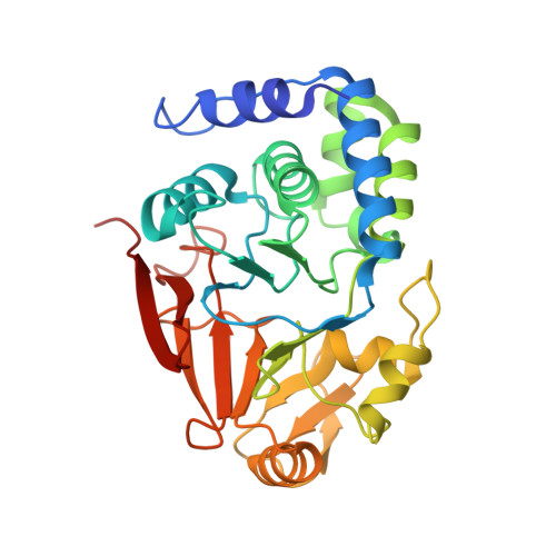
    3N5U

  • PubMed Abstract: 

    The phosphorylation state and corresponding activity of the retinoblastoma tumor suppressor protein (Rb) are modulated by a balance of kinase and phosphatase activities. Here we characterize the association of Rb with the catalytic subunit of protein phosphatase 1 (PP1c). A crystal structure identifies an enzyme docking site in the Rb C-terminal domain that is required for efficient PP1c activity toward Rb. The phosphatase docking site overlaps with the known docking site for cyclin-dependent kinase (Cdk), and PP1 competition with Cdk-cyclins for Rb binding is sufficient to retain Rb activity and block cell-cycle advancement. These results provide the first detailed molecular insights into Rb activation and establish a novel mechanism for Rb regulation in which kinase and phosphatase compete for substrate docking.


  • Organizational Affiliation

    Department of Molecular, Cell, and Developmental Biology, University of California, Santa Cruz, California, USA.


Macromolecules
Find similar proteins by:  (by identity cutoff)  |  3D Structure
Entity ID: 1
MoleculeChains Sequence LengthOrganismDetailsImage
Serine/threonine-protein phosphatase PP1-alpha catalytic subunit
A, B
300Homo sapiensMutation(s): 0 
Gene Names: PPP1APPP1CA
EC: 3.1.3.16
UniProt & NIH Common Fund Data Resources
Find proteins for P62136 (Homo sapiens)
Explore P62136 
Go to UniProtKB:  P62136
PHAROS:  P62136
GTEx:  ENSG00000172531 
Entity Groups  
Sequence Clusters30% Identity50% Identity70% Identity90% Identity95% Identity100% Identity
UniProt GroupP62136
Sequence Annotations
Expand
  • Reference Sequence

Find similar proteins by:  Sequence   |   3D Structure  

Entity ID: 2
MoleculeChains Sequence LengthOrganismDetailsImage
Retinoblastoma-associated protein13Homo sapiensMutation(s): 0 
UniProt & NIH Common Fund Data Resources
Find proteins for P06400 (Homo sapiens)
Explore P06400 
Go to UniProtKB:  P06400
PHAROS:  P06400
GTEx:  ENSG00000139687 
Entity Groups  
Sequence Clusters30% Identity50% Identity70% Identity90% Identity95% Identity100% Identity
UniProt GroupP06400
Sequence Annotations
Expand
  • Reference Sequence
Experimental Data & Validation

Experimental Data

  • Method: X-RAY DIFFRACTION
  • Resolution: 3.20 Å
  • R-Value Free: 0.261 
  • R-Value Work: 0.221 
  • R-Value Observed: 0.225 
  • Space Group: P 41 21 2
Unit Cell:
Length ( Å )Angle ( ˚ )
a = 92.946α = 90
b = 92.946β = 90
c = 192.381γ = 90
Software Package:
Software NamePurpose
MOSFLMdata reduction
SCALAdata scaling
PHASERphasing
PHENIXrefinement
PDB_EXTRACTdata extraction

Structure Validation

View Full Validation Report


Entry History 

Deposition Data

Revision History  (Full details and data files)

  • Version 1.0: 2010-08-11
    Type: Initial release
  • Version 1.1: 2011-07-13
    Changes: Version format compliance
  • Version 1.2: 2024-02-21
    Changes: Data collection, Database references, Derived calculations, Refinement description