3ML4

Crystal structure of a complex between Dok7 PH-PTB and the MuSK juxtamembrane region


Experimental Data Snapshot

  • Method: X-RAY DIFFRACTION
  • Resolution: 2.60 Å
  • R-Value Free: 0.302 
  • R-Value Work: 0.253 
  • R-Value Observed: 0.255 

wwPDB Validation   3D Report Full Report


This is version 1.1 of the entry. See complete history


Literature

The Cytoplasmic Adaptor Protein Dok7 Activates the Receptor Tyrosine Kinase MuSK via Dimerization.

Bergamin, E.Hallock, P.T.Burden, S.J.Hubbard, S.R.

(2010) Mol Cell 39: 100-109

  • DOI: https://doi.org/10.1016/j.molcel.2010.06.007
  • Primary Citation of Related Structures:  
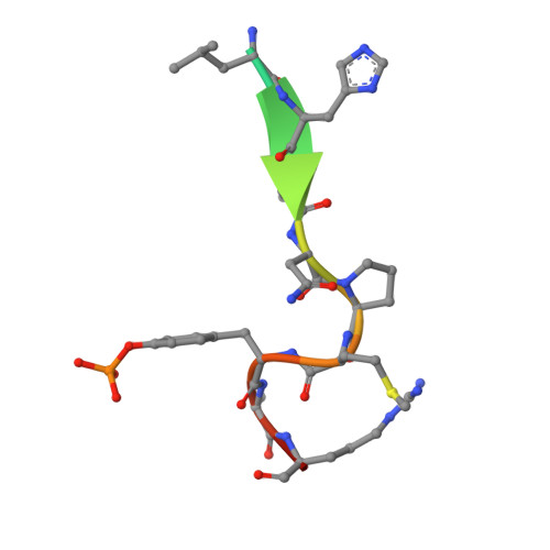
    3ML4

  • PubMed Abstract: 

    Formation of the vertebrate neuromuscular junction requires, among others proteins, Agrin, a neuronally derived ligand, and the following muscle proteins: LRP4, the receptor for Agrin; MuSK, a receptor tyrosine kinase (RTK); and Dok7 (or Dok-7), a cytoplasmic adaptor protein. Dok7 comprises a pleckstrin-homology (PH) domain, a phosphotyrosine-binding (PTB) domain, and C-terminal sites of tyrosine phosphorylation. Unique among adaptor proteins recruited to RTKs, Dok7 is not only a substrate of MuSK, but also an activator of MuSK's kinase activity. Here, we present the crystal structure of the Dok7 PH-PTB domains in complex with a phosphopeptide representing the Dok7-binding site on MuSK. The structure and biochemical data reveal a dimeric arrangement of Dok7 PH-PTB that facilitates trans-autophosphorylation of the kinase activation loop. The structure provides the molecular basis for MuSK activation by Dok7 and for rationalizing several Dok7 loss-of-function mutations found in patients with congenital myasthenic syndromes.


  • Organizational Affiliation

    Structural Biology Program, Kimmel Center for Biology and Medicine of the Skirball Institute and Department of Pharmacology, New York University School of Medicine, New York, NY 10016, USA.


Macromolecules
Find similar proteins by:  (by identity cutoff)  |  3D Structure
Entity ID: 1
MoleculeChains Sequence LengthOrganismDetailsImage
Protein Dok-7
A, B, C, D
224Mus musculusMutation(s): 0 
Gene Names: Dok7
UniProt & NIH Common Fund Data Resources
Find proteins for Q18PE0 (Mus musculus)
Explore Q18PE0 
Go to UniProtKB:  Q18PE0
IMPC:  MGI:3584043
Entity Groups  
Sequence Clusters30% Identity50% Identity70% Identity90% Identity95% Identity100% Identity
UniProt GroupQ18PE0
Sequence Annotations
Expand
  • Reference Sequence

Find similar proteins by:  Sequence   |   3D Structure  

Entity ID: 2
MoleculeChains Sequence LengthOrganismDetailsImage
Muscle, skeletal receptor tyrosine-protein kinase
E, F, G, H
13Mus musculusMutation(s): 0 
UniProt
Find proteins for Q61006 (Mus musculus)
Explore Q61006 
Go to UniProtKB:  Q61006
Entity Groups  
Sequence Clusters30% Identity50% Identity70% Identity90% Identity95% Identity100% Identity
UniProt GroupQ61006
Sequence Annotations
Expand
  • Reference Sequence
Small Molecules
Modified Residues  2 Unique
IDChains TypeFormula2D DiagramParent
MSE
Query on MSE
A, B, C, D
L-PEPTIDE LINKINGC5 H11 N O2 SeMET
PTR
Query on PTR
E, F, G, H
L-PEPTIDE LINKINGC9 H12 N O6 PTYR
Experimental Data & Validation

Experimental Data

  • Method: X-RAY DIFFRACTION
  • Resolution: 2.60 Å
  • R-Value Free: 0.302 
  • R-Value Work: 0.253 
  • R-Value Observed: 0.255 
  • Space Group: P 32 2 1
Unit Cell:
Length ( Å )Angle ( ˚ )
a = 134.6α = 90
b = 134.6β = 90
c = 121.58γ = 120
Software Package:
Software NamePurpose
REFMACrefinement

Structure Validation

View Full Validation Report


Entry History 

Deposition Data

Revision History  (Full details and data files)

  • Version 1.0: 2010-07-14
    Type: Initial release
  • Version 1.1: 2011-07-13
    Changes: Version format compliance