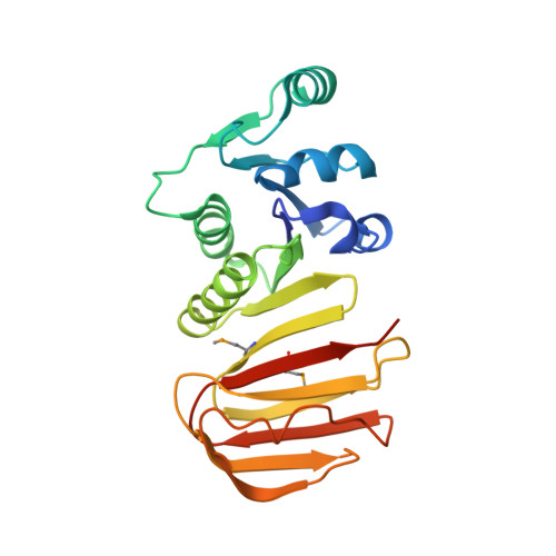
Northeast Structural Genomics Consortium Target SR677
Kuzin, A., Abashidze, M., Seetharaman, J., Mao, M., Xiao, R., Foote, E.L., Ciccosanti, C., Wang, H., Everett, J.K., Nair, R., Acton, T.B., Rost, B., Montelione, G.T., Hunt, J.F., Tong, L.To be published.