3HRF

Crystal structure of Human PDK1 kinase domain in complex with an allosteric activator bound to the PIF-pocket


Experimental Data Snapshot

  • Method: X-RAY DIFFRACTION
  • Resolution: 1.90 Å
  • R-Value Free: 0.177 
  • R-Value Work: 0.165 
  • R-Value Observed: 0.165 

wwPDB Validation   3D Report Full Report


Ligand Structure Quality Assessment 


This is version 1.4 of the entry. See complete history


Literature

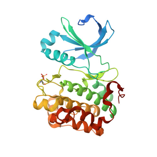
Structure and allosteric effects of low-molecular-weight activators on the protein kinase PDK1.

Hindie, V.Stroba, A.Zhang, H.Lopez-Garcia, L.A.Idrissova, L.Zeuzem, S.Hirschberg, D.Schaeffer, F.Jrgensen, T.J.Engel, M.Alzari, P.M.Biondi, R.M.

(2009) Nat Chem Biol 5: 758-764

  • DOI: https://doi.org/10.1038/nchembio.208
  • Primary Citation of Related Structures:  
    3HRC, 3HRF

  • PubMed Abstract: 

    Protein phosphorylation transduces a large set of intracellular signals. One mechanism by which phosphorylation mediates signal transduction is by prompting conformational changes in the target protein or interacting proteins. Previous work described an allosteric site mediating phosphorylation-dependent activation of AGC kinases. The AGC kinase PDK1 is activated by the docking of a phosphorylated motif from substrates. Here we present the crystallography of PDK1 bound to a rationally developed low-molecular-weight activator and describe the conformational changes induced by small compounds in the crystal and in solution using a fluorescence-based assay and deuterium exchange experiments. Our results indicate that the binding of the compound produces local changes at the target site, the PIF binding pocket, and also allosteric changes at the ATP binding site and the activation loop. Altogether, we present molecular details of the allosteric changes induced by small compounds that trigger the activation of PDK1 through mimicry of phosphorylation-dependent conformational changes.


  • Organizational Affiliation

    Research Group PhosphoSites, Department of Internal Medicine I, Universitätsklinikum Frankfurt, Frankfurt, Germany.


Macromolecules
Find similar proteins by:  (by identity cutoff)  |  3D Structure
Entity ID: 1
MoleculeChains Sequence LengthOrganismDetailsImage
3-phosphoinositide-dependent protein kinase 1311Homo sapiensMutation(s): 2 
Gene Names: PDK1PDPK1
UniProt & NIH Common Fund Data Resources
Find proteins for O15530 (Homo sapiens)
Explore O15530 
Go to UniProtKB:  O15530
PHAROS:  O15530
GTEx:  ENSG00000140992 
Entity Groups  
Sequence Clusters30% Identity50% Identity70% Identity90% Identity95% Identity100% Identity
UniProt GroupO15530
Sequence Annotations
Expand
  • Reference Sequence
Small Molecules
Ligands 3 Unique
IDChains Name / Formula / InChI Key2D Diagram3D Interactions
ATP
Query on ATP

Download Ideal Coordinates CCD File 
B [auth A]ADENOSINE-5'-TRIPHOSPHATE
C10 H16 N5 O13 P3
ZKHQWZAMYRWXGA-KQYNXXCUSA-N
P47
Query on P47

Download Ideal Coordinates CCD File 
C [auth A](2Z)-5-(4-chlorophenyl)-3-phenylpent-2-enoic acid
C17 H15 Cl O2
LLJYFDRQFPQGNY-QINSGFPZSA-N
EPE
Query on EPE

Download Ideal Coordinates CCD File 
D [auth A],
E [auth A]
4-(2-HYDROXYETHYL)-1-PIPERAZINE ETHANESULFONIC ACID
C8 H18 N2 O4 S
JKMHFZQWWAIEOD-UHFFFAOYSA-N
Modified Residues  1 Unique
IDChains TypeFormula2D DiagramParent
SEP
Query on SEP
A
L-PEPTIDE LINKINGC3 H8 N O6 PSER
Binding Affinity Annotations 
IDSourceBinding Affinity
P47 BindingDB:  3HRF Kd: 1.03e+4 (nM) from 1 assay(s)
IC50: 9.70e+4 (nM) from 1 assay(s)
Binding MOAD:  3HRF Kd: 1.03e+4 (nM) from 1 assay(s)
PDBBind:  3HRF Kd: 1.03e+4 (nM) from 1 assay(s)
Experimental Data & Validation

Experimental Data

  • Method: X-RAY DIFFRACTION
  • Resolution: 1.90 Å
  • R-Value Free: 0.177 
  • R-Value Work: 0.165 
  • R-Value Observed: 0.165 
  • Space Group: C 1 2 1
Unit Cell:
Length ( Å )Angle ( ˚ )
a = 148.55α = 90
b = 44β = 100.42
c = 47.24γ = 90
Software Package:
Software NamePurpose
REFMACrefinement
PDB_EXTRACTdata extraction
XDSdata scaling
XDSdata reduction
SCALAdata scaling
PHASERphasing

Structure Validation

View Full Validation Report


Ligand Structure Quality Assessment 


Entry History 

Deposition Data

Revision History  (Full details and data files)

  • Version 1.0: 2009-09-15
    Type: Initial release
  • Version 1.1: 2011-07-13
    Changes: Advisory, Refinement description, Version format compliance
  • Version 1.2: 2013-11-13
    Changes: Database references
  • Version 1.3: 2021-11-10
    Changes: Database references, Derived calculations
  • Version 1.4: 2023-11-01
    Changes: Data collection, Refinement description