3H1D

Structure of the HUWE1 HECT Domain


Experimental Data Snapshot

  • Method: X-RAY DIFFRACTION
  • Resolution: 1.89 Å
  • R-Value Free: 0.229 
  • R-Value Work: 0.166 
  • R-Value Observed: 0.169 

wwPDB Validation   3D Report Full Report


This is version 1.3 of the entry. See complete history


Literature

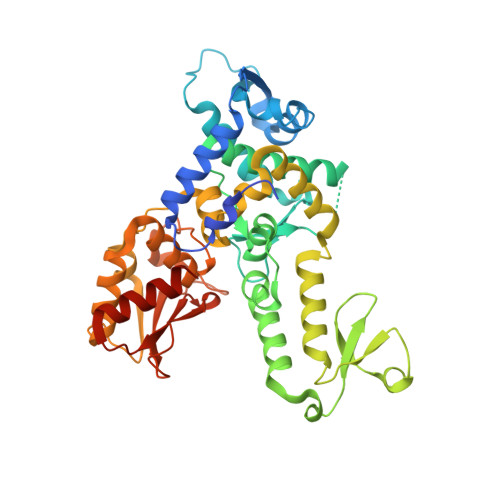
A structural element within the HUWE1 HECT domain modulates self-ubiquitination and substrate ubiquitination activities.

Pandya, R.K.Partridge, J.R.Love, K.R.Schwartz, T.U.Ploegh, H.L.

(2010) J Biol Chem 285: 5664-5673

  • DOI: https://doi.org/10.1074/jbc.M109.051805
  • Primary Citation of Related Structures:  
    3H1D

  • PubMed Abstract: 

    E3 ubiquitin ligases catalyze the final step of ubiquitin conjugation and regulate numerous cellular processes. The HECT class of E3 ubiquitin (Ub) ligases directly transfers Ub from bound E2 enzyme to a myriad of substrates. The catalytic domain of HECT Ub ligases has a bilobal architecture that separates the E2 binding region and catalytic site. An important question regarding HECT domain function is the control of ligase activity and specificity. Here we present a functional analysis of the HECT domain of the E3 ligase HUWE1 based on crystal structures and show that a single N-terminal helix significantly stabilizes the HECT domain. We observe that this element modulates HECT domain activity, as measured by self-ubiquitination induced in the absence of this helix, as distinct from its effects on Ub conjugation of substrate Mcl-1. Such subtle changes to the protein may be at the heart of the vast spectrum of substrate specificities displayed by HECT domain E3 ligases.


  • Organizational Affiliation

    Whitehead Institute for Biomedical Research, Cambridge, Massachusetts 02142, USA.


Macromolecules
Find similar proteins by:  (by identity cutoff)  |  3D Structure
Entity ID: 1
MoleculeChains Sequence LengthOrganismDetailsImage
E3 ubiquitin-protein ligase HUWE1405Homo sapiensMutation(s): 3 
Gene Names: HUWE1KIAA0312KIAA1578UREB1HSPC272
EC: 6.3.2
UniProt & NIH Common Fund Data Resources
Find proteins for Q7Z6Z7 (Homo sapiens)
Explore Q7Z6Z7 
Go to UniProtKB:  Q7Z6Z7
PHAROS:  Q7Z6Z7
GTEx:  ENSG00000086758 
Entity Groups  
Sequence Clusters30% Identity50% Identity70% Identity90% Identity95% Identity100% Identity
UniProt GroupQ7Z6Z7
Sequence Annotations
Expand
  • Reference Sequence
Experimental Data & Validation

Experimental Data

  • Method: X-RAY DIFFRACTION
  • Resolution: 1.89 Å
  • R-Value Free: 0.229 
  • R-Value Work: 0.166 
  • R-Value Observed: 0.169 
  • Space Group: C 1 2 1
Unit Cell:
Length ( Å )Angle ( ˚ )
a = 119.654α = 90
b = 55.653β = 122.49
c = 69.585γ = 90
Software Package:
Software NamePurpose
ADSCdata collection
PHASERphasing
PHENIXrefinement
HKL-2000data reduction
SCALEPACKdata scaling

Structure Validation

View Full Validation Report


Entry History 

Deposition Data

Revision History  (Full details and data files)

  • Version 1.0: 2009-12-08
    Type: Initial release
  • Version 1.1: 2011-07-13
    Changes: Version format compliance
  • Version 1.2: 2021-10-13
    Changes: Database references, Derived calculations
  • Version 1.3: 2023-09-06
    Changes: Data collection, Refinement description