3G76

Crystal structure of XIAP-BIR3 in complex with a bivalent compound


Experimental Data Snapshot

  • Method: X-RAY DIFFRACTION
  • Resolution: 3.00 Å
  • R-Value Free: 0.311 
  • R-Value Work: 0.231 
  • R-Value Observed: 0.235 

wwPDB Validation   3D Report Full Report


This is version 1.2 of the entry. See complete history


Literature

Structural basis for bivalent smac-mimetics recognition in the IAP protein family

Cossu, F.Milani, M.Mastrangelo, E.Vachette, P.Servida, F.Lecis, D.Canevari, G.Delia, D.Drago, C.Rizzo, V.Manzoni, L.Seneci, P.Scolastico, C.Bolognesi, M.

(2009) J Mol Biol 392: 630-644

  • DOI: https://doi.org/10.1016/j.jmb.2009.04.033
  • Primary Citation of Related Structures:  
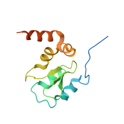
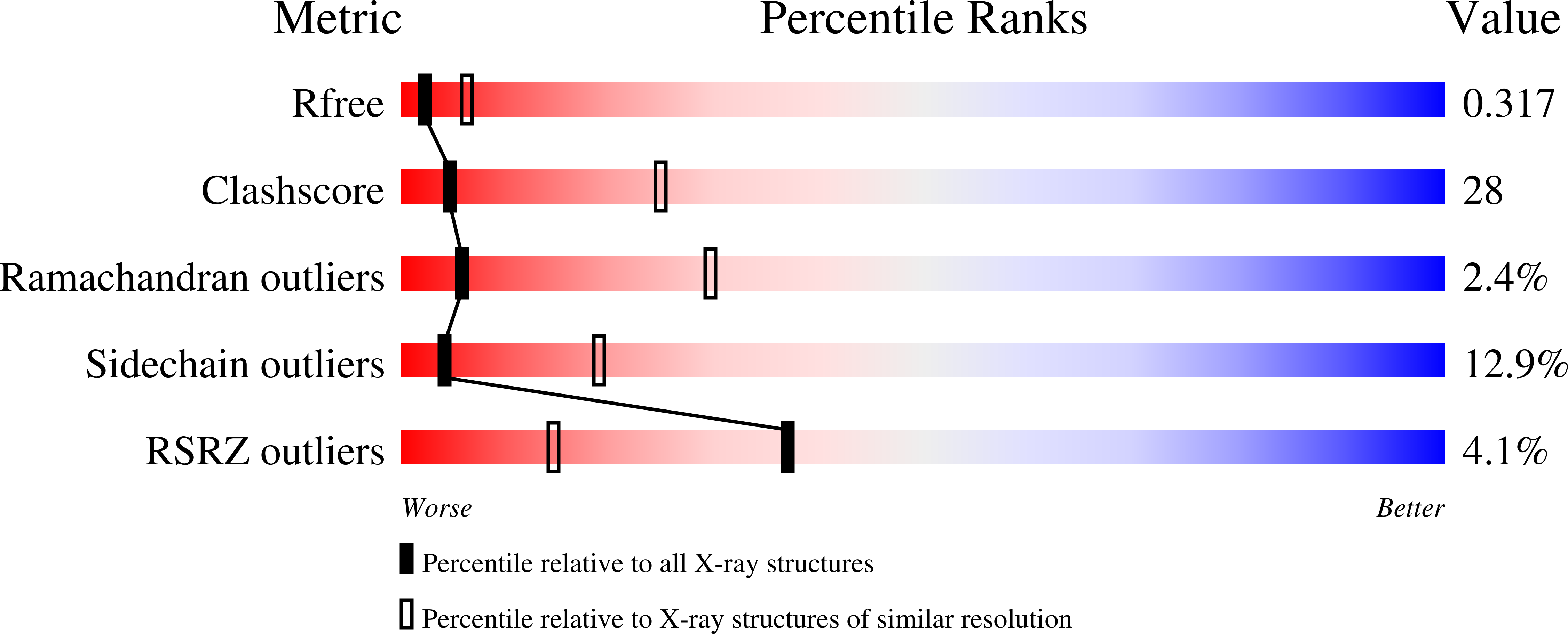
    3G76

  • PubMed Abstract: 

    XIAP is an apoptotic regulator protein that binds to the effector caspases -3 and -7 through its BIR2 domain, and to initiator caspase-9 through its BIR3 domain. Molecular docking studies suggested that Smac-DIABLO may antagonize XIAP by concurrently targeting both BIR2 and BIR3 domains; on this basis bivalent Smac-mimetic compounds have been proposed and characterized. Here, we report the X-ray crystal structure of XIAP-BIR3 domain in complex with a two-headed compound (compound 3) with improved efficacy relative to its monomeric form. A small-angle X-ray scattering study of XIAP-BIR2BIR3, together with fluorescence polarization binding assays and compound 3 cytotoxicity tests on HL60 leukemia cell line are also reported. The crystal structure analysis reveals a network of interactions supporting XIAP-BIR3/compound 3 recognition; moreover, analytical gel-filtration chromatography shows that compound 3 forms a 1:1 stoichiometric complex with a XIAP protein construct containing both BIR2 and BIR3 domains. On the basis of the crystal structure and small-angle X-ray scattering, a model of the same BIR2-BIR3 construct bound to compound 3 is proposed, shedding light on the ability of compound 3 to relieve XIAP inhibitory effects on caspase-9 as well as caspases -3 and -7. A molecular modeling/docking analysis of compound 3 bound to cIAP1-BIR3 domain is presented, considering that Smac-mimetics have been shown to kill tumor cells by inducing cIAP1 and cIAP2 ubiquitination and degradation. Taken together, the results reported here provide a rationale for further development of compound 3 as a lead in the design of dimeric Smac mimetics for cancer treatment.


  • Organizational Affiliation

    Department of Biomolecular Sciences and Biotechnology, University of Milano, Italy.


Macromolecules
Find similar proteins by:  (by identity cutoff)  |  3D Structure
Entity ID: 1
MoleculeChains Sequence LengthOrganismDetailsImage
Baculoviral IAP repeat-containing protein 4
A, B, C, D, E
A, B, C, D, E, F, G, H
122Homo sapiensMutation(s): 0 
Gene Names: XIAP
EC: 6.3.2
UniProt & NIH Common Fund Data Resources
Find proteins for P98170 (Homo sapiens)
Explore P98170 
Go to UniProtKB:  P98170
PHAROS:  P98170
GTEx:  ENSG00000101966 
Entity Groups  
Sequence Clusters30% Identity50% Identity70% Identity90% Identity95% Identity100% Identity
UniProt GroupP98170
Sequence Annotations
Expand
  • Reference Sequence
Small Molecules
Ligands 2 Unique
IDChains Name / Formula / InChI Key2D Diagram3D Interactions
CZ3
Query on CZ3

Download Ideal Coordinates CCD File 
J [auth A]
L [auth B]
N [auth C]
P [auth D]
R [auth E]
J [auth A],
L [auth B],
N [auth C],
P [auth D],
R [auth E],
T [auth F],
V [auth G],
X [auth H]
1,1'-{hexa-2,4-diyne-1,6-diylbis[oxy{(2S,3R)-2-[(N-methyl-L-alanyl)amino]-1-oxobutane-3,1-diyl}(2S)pyrrolidine-1,2-diylmethanediyl]}bis[5-(phenylsulfanyl)-1H-tetrazole]
C46 H60 N14 O6 S2
CDUYVSXKBIKUNH-RAUIGOKHSA-N
ZN
Query on ZN

Download Ideal Coordinates CCD File 
I [auth A]
K [auth B]
M [auth C]
O [auth D]
Q [auth E]
I [auth A],
K [auth B],
M [auth C],
O [auth D],
Q [auth E],
S [auth F],
U [auth G],
W [auth H]
ZINC ION
Zn
PTFCDOFLOPIGGS-UHFFFAOYSA-N
Binding Affinity Annotations 
IDSourceBinding Affinity
CZ3 PDBBind:  3G76 IC50: 230.8 (nM) from 1 assay(s)
Experimental Data & Validation

Experimental Data

  • Method: X-RAY DIFFRACTION
  • Resolution: 3.00 Å
  • R-Value Free: 0.311 
  • R-Value Work: 0.231 
  • R-Value Observed: 0.235 
  • Space Group: P 31
Unit Cell:
Length ( Å )Angle ( ˚ )
a = 119.077α = 90
b = 119.077β = 90
c = 105.59γ = 120
Software Package:
Software NamePurpose
REFMACrefinement
MOSFLMdata reduction
SCALAdata scaling

Structure Validation

View Full Validation Report


Entry History 

Deposition Data

Revision History  (Full details and data files)

  • Version 1.0: 2009-05-12
    Type: Initial release
  • Version 1.1: 2011-07-13
    Changes: Version format compliance
  • Version 1.2: 2023-11-01
    Changes: Data collection, Database references, Derived calculations, Refinement description