


3FAC
Crystal structure of Rhodobacter sphaeroides protein RSP_2168. Northeast Structural Genomics target RhR83.
- PDB DOI: https://doi.org/10.2210/pdb3FAC/pdb
- Classification: UNKNOWN FUNCTION
- Organism(s): Cereibacter sphaeroides 2.4.1
- Expression System: Escherichia coli
- Mutation(s): No
- Deposited: 2008-11-16 Released: 2008-12-09
Experimental Data Snapshot
- Method: X-RAY DIFFRACTION
- Resolution: 2.50 Å
- R-Value Free: 0.258
- R-Value Work: 0.225
- R-Value Observed: 0.225
wwPDB Validation 3D Report Full Report

This is version 1.2 of the entry. See complete history.

Macromolecules
Find similar proteins by:
(by identity cutoff) | 3D Structure
Entity ID: 1 | |||||
|---|---|---|---|---|---|
| Molecule | Chains | Sequence Length | Organism | Details | Image |
| Putative uncharacterized protein | 118 | Cereibacter sphaeroides 2.4.1 | Mutation(s): 0 Gene Names: RHOS4_07510, RSP_2168 | 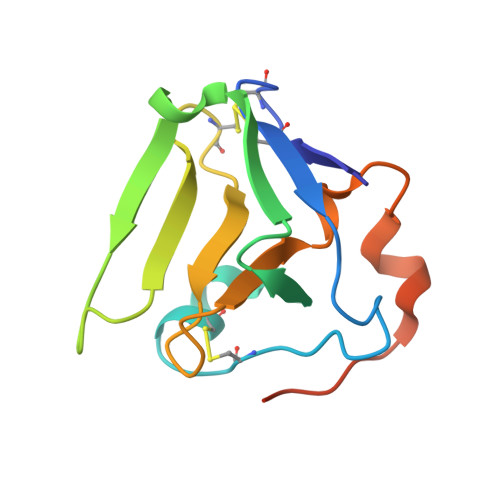 | |
UniProt | |||||
Find proteins for Q3J4G5 (Cereibacter sphaeroides (strain ATCC 17023 / DSM 158 / JCM 6121 / CCUG 31486 / LMG 2827 / NBRC 12203 / NCIMB 8253 / ATH 2.4.1.)) Explore Q3J4G5 Go to UniProtKB: Q3J4G5 | |||||
Entity Groups | |||||
| Sequence Clusters | 30% Identity50% Identity70% Identity90% Identity95% Identity100% Identity | ||||
| UniProt Group | Q3J4G5 | ||||
Sequence AnnotationsExpand | |||||
| |||||
Experimental Data & Validation
Experimental Data
- Method: X-RAY DIFFRACTION
- Resolution: 2.50 Å
- R-Value Free: 0.258
- R-Value Work: 0.225
- R-Value Observed: 0.225
- Space Group: P 21 21 21
Unit Cell:
| Length ( Å ) | Angle ( ˚ ) |
|---|---|
| a = 68.579 | α = 90 |
| b = 125.996 | β = 90 |
| c = 135.791 | γ = 90 |
| Software Name | Purpose |
|---|---|
| ADSC | data collection |
| SOLVE | phasing |
| CNS | refinement |
| HKL-2000 | data reduction |
| HKL-2000 | data scaling |
Entry History
Deposition Data
- Released Date: 2008-12-09 Deposition Author(s): Seetharaman, J., Neely, H., Forouhar, F., Wang, H., Janjua, H., Foote, E.L., Xiao, R., Everett, J.K., Acton, T.B., Rost, B., Montelione, G.T., Hunt, J.F., Tong, L., Northeast Structural Genomics Consortium (NESG)
Revision History (Full details and data files)
- Version 1.0: 2008-12-09
Type: Initial release - Version 1.1: 2011-07-13
Changes: Version format compliance - Version 1.2: 2023-12-27
Changes: Data collection, Database references
