3F69

Crystal structure of the Mycobacterium tuberculosis PknB mutant kinase domain in complex with KT5720


Experimental Data Snapshot

  • Method: X-RAY DIFFRACTION
  • Resolution: 2.80 Å
  • R-Value Free: 0.268 
  • R-Value Work: 0.218 
  • R-Value Observed: 0.221 

wwPDB Validation   3D Report Full Report


Ligand Structure Quality Assessment 


This is version 1.5 of the entry. See complete history


Literature

Auto-activation mechanism of the Mycobacterium tuberculosis PknB receptor Ser/Thr kinase.

Mieczkowski, C.Iavarone, A.T.Alber, T.

(2008) EMBO J 27: 3186-3197

  • DOI: https://doi.org/10.1038/emboj.2008.236
  • Primary Citation of Related Structures:  
    3F61, 3F69

  • PubMed Abstract: 

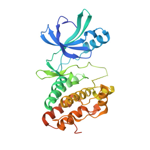
    Many Ser/Thr protein kinases are activated by autophosphorylation, but the mechanism of this process has not been defined. We determined the crystal structure of a mutant of the Ser/Thr kinase domain (KD) of the mycobacterial sensor kinase PknB in complex with an ATP competitive inhibitor and discovered features consistent with an activation complex. The complex formed an asymmetric dimer, with the G helix and the ordered activation loop of one KD in contact with the G helix of the other. The activation loop of this putative 'substrate' KD was disordered, with the ends positioned at the entrance to the partner KD active site. Single amino-acid substitutions in the G-helix interface reduced activation-loop phosphorylation, and multiple replacements abolished KD phosphorylation and kinase activation. Phosphorylation of an inactive mutant KD was reduced by G-helix substitutions in both active and inactive KDs, as predicted by the idea that the asymmetric dimer mimics a trans-autophosphorylation complex. These results support a model in which a structurally and functionally asymmetric, 'front-to-front' association mediates autophosphorylation of PknB and homologous kinases.


  • Organizational Affiliation

    Department of Molecular and Cell Biology, University of California, Berkeley, CA 94720-3220, USA.


Macromolecules
Find similar proteins by:  (by identity cutoff)  |  3D Structure
Entity ID: 1
MoleculeChains Sequence LengthOrganismDetailsImage
Serine/threonine-protein kinase pknB
A, B
311Mycobacterium tuberculosisMutation(s): 3 
Gene Names: MT0017MTCY10H4.14cpknBRv0014c
EC: 2.7.11.1
UniProt
Find proteins for P9WI81 (Mycobacterium tuberculosis (strain ATCC 25618 / H37Rv))
Explore P9WI81 
Go to UniProtKB:  P9WI81
Entity Groups  
Sequence Clusters30% Identity50% Identity70% Identity90% Identity95% Identity100% Identity
UniProt GroupP9WI81
Sequence Annotations
Expand
  • Reference Sequence
Small Molecules
Modified Residues  1 Unique
IDChains TypeFormula2D DiagramParent
TPO
Query on TPO
A, B
L-PEPTIDE LINKINGC4 H10 N O6 PTHR
Binding Affinity Annotations 
IDSourceBinding Affinity
XDR PDBBind:  3F69 IC50: 1000 (nM) from 1 assay(s)
Experimental Data & Validation

Experimental Data

  • Method: X-RAY DIFFRACTION
  • Resolution: 2.80 Å
  • R-Value Free: 0.268 
  • R-Value Work: 0.218 
  • R-Value Observed: 0.221 
  • Space Group: F 41 3 2
Unit Cell:
Length ( Å )Angle ( ˚ )
a = 297.557α = 90
b = 297.557β = 90
c = 297.557γ = 90
Software Package:
Software NamePurpose
DENZOdata reduction
SCALEPACKdata scaling
PHASERphasing
REFMACrefinement
PDB_EXTRACTdata extraction
HKL-2000data collection

Structure Validation

View Full Validation Report


Ligand Structure Quality Assessment 


Entry History 

Deposition Data

Revision History  (Full details and data files)

  • Version 1.0: 2008-12-02
    Type: Initial release
  • Version 1.1: 2011-07-13
    Changes: Advisory, Refinement description, Version format compliance
  • Version 1.2: 2017-10-25
    Changes: Advisory, Refinement description
  • Version 1.3: 2021-10-20
    Changes: Advisory, Database references, Derived calculations, Structure summary
  • Version 1.4: 2023-12-27
    Changes: Data collection
  • Version 1.5: 2024-04-03
    Changes: Refinement description