3EFK

Structure of c-Met with pyrimidone inhibitor 50


Experimental Data Snapshot

  • Method: X-RAY DIFFRACTION
  • Resolution: 2.20 Å
  • R-Value Free: 0.292 
  • R-Value Work: 0.256 
  • R-Value Observed: 0.257 

wwPDB Validation   3D Report Full Report


Ligand Structure Quality Assessment 


This is version 1.3 of the entry. See complete history


Literature

Design, synthesis, and biological evaluation of potent c-Met inhibitors.

D'Angelo, N.D.Bellon, S.F.Booker, S.K.Cheng, Y.Coxon, A.Dominguez, C.Fellows, I.Hoffman, D.Hungate, R.Kaplan-Lefko, P.Lee, M.R.Li, C.Liu, L.Rainbeau, E.Reider, P.J.Rex, K.Siegmund, A.Sun, Y.Tasker, A.S.Xi, N.Xu, S.Yang, Y.Zhang, Y.Burgess, T.L.Dussault, I.Kim, T.S.

(2008) J Med Chem 51: 5766-5779

  • DOI: https://doi.org/10.1021/jm8006189
  • Primary Citation of Related Structures:  
    3EFJ, 3EFK

  • PubMed Abstract: 

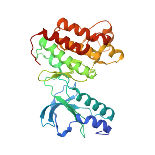
    c-Met is a receptor tyrosine kinase that plays a key role in several cellular processes but has also been found to be overexpressed and mutated in different human cancers. Consequently, targeting this enzyme has become an area of intense research in drug discovery. Our studies began with the design and synthesis of novel pyrimidone 7, which was found to be a potent c-Met inhibitor. Subsequent SAR studies identified 22 as a more potent analog, whereas an X-ray crystal structure of 7 bound to c-Met revealed an unexpected binding conformation. This latter finding led to the development of a new series that featured compounds that were more potent both in vitro and in vivo than 22 and also exhibited different binding conformations to c-Met. Novel c-Met inhibitors have been designed, developed, and found to be potent in vitro and in vivo.


  • Organizational Affiliation

    Department of Medicinal Chemistry, Amgen Inc., One Amgen Center Drive, Thousand Oaks, California 91320-1799, USA. dangelo@amgen.com


Macromolecules
Find similar proteins by:  (by identity cutoff)  |  3D Structure
Entity ID: 1
MoleculeChains Sequence LengthOrganismDetailsImage
Hepatocyte growth factor receptor
A, B
310Homo sapiensMutation(s): 1 
Gene Names: MET
EC: 2.7.10.1
UniProt & NIH Common Fund Data Resources
Find proteins for P08581 (Homo sapiens)
Explore P08581 
Go to UniProtKB:  P08581
PHAROS:  P08581
GTEx:  ENSG00000105976 
Entity Groups  
Sequence Clusters30% Identity50% Identity70% Identity90% Identity95% Identity100% Identity
UniProt GroupP08581
Sequence Annotations
Expand
  • Reference Sequence
Small Molecules
Ligands 1 Unique
IDChains Name / Formula / InChI Key2D Diagram3D Interactions
MT4
Query on MT4

Download Ideal Coordinates CCD File 
C [auth A],
D [auth B]
5-{4-[(6,7-dimethoxyquinolin-4-yl)oxy]-3-fluorophenyl}-2-[(4-fluorophenyl)amino]-3-methylpyrimidin-4(3H)-one
C28 H22 F2 N4 O4
DUTNABYDBFVZOA-UHFFFAOYSA-N
Binding Affinity Annotations 
IDSourceBinding Affinity
MT4 BindingDB:  3EFK Ki: 14 (nM) from 1 assay(s)
IC50: 14 (nM) from 1 assay(s)
PDBBind:  3EFK Ki: 14 (nM) from 1 assay(s)
Binding MOAD:  3EFK Ki: 14 (nM) from 1 assay(s)
Experimental Data & Validation

Experimental Data

  • Method: X-RAY DIFFRACTION
  • Resolution: 2.20 Å
  • R-Value Free: 0.292 
  • R-Value Work: 0.256 
  • R-Value Observed: 0.257 
  • Space Group: P 21 21 21
Unit Cell:
Length ( Å )Angle ( ˚ )
a = 76.205α = 90
b = 78.25β = 90
c = 129.684γ = 90
Software Package:
Software NamePurpose
CrystalCleardata collection
AMoREphasing
REFMACrefinement
DENZOdata reduction
SCALEPACKdata scaling

Structure Validation

View Full Validation Report


Ligand Structure Quality Assessment 


Entry History 

Deposition Data

Revision History  (Full details and data files)

  • Version 1.0: 2008-10-07
    Type: Initial release
  • Version 1.1: 2011-07-13
    Changes: Version format compliance
  • Version 1.2: 2021-10-20
    Changes: Database references, Derived calculations
  • Version 1.3: 2023-08-30
    Changes: Data collection, Refinement description