3BRB

Crystal structure of catalytic domain of the proto-oncogene tyrosine-protein kinase MER in complex with ADP


Experimental Data Snapshot

  • Method: X-RAY DIFFRACTION
  • Resolution: 1.90 Å
  • R-Value Free: 0.240 
  • R-Value Work: 0.192 
  • R-Value Observed: 0.195 

wwPDB Validation   3D Report Full Report


Ligand Structure Quality Assessment 


This is version 1.2 of the entry. See complete history


Literature

Structural insights into the inhibited states of the Mer receptor tyrosine kinase.

Huang, X.Finerty, P.Walker, J.R.Butler-Cole, C.Vedadi, M.Schapira, M.Parker, S.A.Turk, B.E.Thompson, D.A.Dhe-Paganon, S.

(2009) J Struct Biol 165: 88-96

  • DOI: https://doi.org/10.1016/j.jsb.2008.10.003
  • Primary Citation of Related Structures:  
    2P0C, 3BPR, 3BRB

  • PubMed Abstract: 

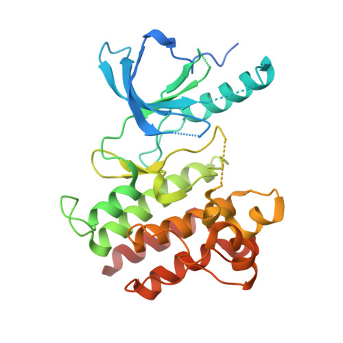
    The mammalian ortholog of the retroviral oncogene v-Eyk, and a receptor tyrosine kinase upstream of antiapoptotic and transforming signals, Mer (MerTK) is a mediator of the phagocytic process, being involved in retinal and immune cell clearance and platelet aggregation. Mer knockout mice are viable and are protected from epinephrine-induced pulmonary thromboembolism and ferric chloride-induced thrombosis. Mer overexpression, on the other hand, is associated with numerous carcinomas. Although Mer adaptor proteins and signaling pathways have been identified, it remains unclear how Mer initiates phagocytosis. When bound to its nucleotide cofactor, the high-resolution structure of Mer shows an autoinhibited alphaC-Glu-out conformation with insertion of an activation loop residue into the active site. Mer complexed with compound-52 (C52: 2-(2-hydroxyethylamino)-6-(3-chloroanilino)-9-isopropylpurine), a ligand identified from a focused library, retains its DFG-Asp-in and alphaC-Glu-out conformation, but acquires other conformational changes. The alphaC helix and DFGL region is closer to the hinge region and the ethanolamine moiety of C52 binds in the groove formed between Leu593 and Val601 of the P-loop, causing a compression of the active site pocket. These conformational states reveal the mechanisms of autoinhibition, the pathophysiological basis of disease-causing mutations, and a platform for the development of chemical probes.


  • Organizational Affiliation

    Structural Genomics Consortium, University of Toronto, Ont., Canada.


Macromolecules
Find similar proteins by:  (by identity cutoff)  |  3D Structure
Entity ID: 1
MoleculeChains Sequence LengthOrganismDetailsImage
Proto-oncogene tyrosine-protein kinase MER
A, B
313Homo sapiensMutation(s): 0 
Gene Names: MERTKMER
EC: 2.7.10.1
UniProt & NIH Common Fund Data Resources
Find proteins for Q12866 (Homo sapiens)
Explore Q12866 
Go to UniProtKB:  Q12866
PHAROS:  Q12866
GTEx:  ENSG00000153208 
Entity Groups  
Sequence Clusters30% Identity50% Identity70% Identity90% Identity95% Identity100% Identity
UniProt GroupQ12866
Sequence Annotations
Expand
  • Reference Sequence
Small Molecules
Ligands 4 Unique
IDChains Name / Formula / InChI Key2D Diagram3D Interactions
ADP
Query on ADP

Download Ideal Coordinates CCD File 
F [auth A],
L [auth B]
ADENOSINE-5'-DIPHOSPHATE
C10 H15 N5 O10 P2
XTWYTFMLZFPYCI-KQYNXXCUSA-N
GOL
Query on GOL

Download Ideal Coordinates CCD File 
G [auth A]GLYCEROL
C3 H8 O3
PEDCQBHIVMGVHV-UHFFFAOYSA-N
CL
Query on CL

Download Ideal Coordinates CCD File 
C [auth A],
D [auth A],
H [auth B],
I [auth B],
J [auth B]
CHLORIDE ION
Cl
VEXZGXHMUGYJMC-UHFFFAOYSA-M
MG
Query on MG

Download Ideal Coordinates CCD File 
E [auth A],
K [auth B]
MAGNESIUM ION
Mg
JLVVSXFLKOJNIY-UHFFFAOYSA-N
Experimental Data & Validation

Experimental Data

  • Method: X-RAY DIFFRACTION
  • Resolution: 1.90 Å
  • R-Value Free: 0.240 
  • R-Value Work: 0.192 
  • R-Value Observed: 0.195 
  • Space Group: P 1 21 1
Unit Cell:
Length ( Å )Angle ( ˚ )
a = 53.385α = 90
b = 89.88β = 103.09
c = 69.537γ = 90
Software Package:
Software NamePurpose
PHASERphasing
REFMACrefinement
HKL-2000data collection
HKL-2000data reduction
HKL-2000data scaling

Structure Validation

View Full Validation Report


Ligand Structure Quality Assessment 


Entry History 

Revision History  (Full details and data files)

  • Version 1.0: 2008-01-22
    Type: Initial release
  • Version 1.1: 2011-07-13
    Changes: Non-polymer description, Version format compliance
  • Version 1.2: 2023-08-30
    Changes: Data collection, Database references, Derived calculations, Refinement description