


X-Ray structure of the P64488 from E.coli (strain K12).
Kuzin, A.P., Su, M., Seetharaman, J., Wang, D., Janjua, H., Owens, L., Ma, L.-C., Xiao, R., Liu, J., Baran, M.C., Acton, T.B., Rost, B., Montelione, G.T., Tong, L., Hunt, J.F.To be published.
Experimental Data Snapshot
wwPDB Validation 3D Report Full Report

Entity ID: 1 | |||||
|---|---|---|---|---|---|
| Molecule | Chains | Sequence Length | Organism | Details | Image |
| Uncharacterized protein yeaR | 127 | Escherichia coli K-12 | Mutation(s): 0 Gene Names: yeaR, b1797, JW1786 | 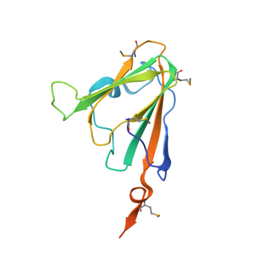 | |
UniProt | |||||
Find proteins for P64488 (Escherichia coli (strain K12)) Explore P64488 Go to UniProtKB: P64488 | |||||
Entity Groups | |||||
| Sequence Clusters | 30% Identity50% Identity70% Identity90% Identity95% Identity100% Identity | ||||
| UniProt Group | P64488 | ||||
Sequence AnnotationsExpand | |||||
| |||||
| Ligands 1 Unique | |||||
|---|---|---|---|---|---|
| ID | Chains | Name / Formula / InChI Key | 2D Diagram | 3D Interactions | |
| ZN Query on ZN | E [auth A], F [auth B], G [auth C], H [auth D] | ZINC ION Zn PTFCDOFLOPIGGS-UHFFFAOYSA-N |  | ||
| Modified Residues 1 Unique | |||||
|---|---|---|---|---|---|
| ID | Chains | Type | Formula | 2D Diagram | Parent |
| MSE Query on MSE | A, B, C, D | L-PEPTIDE LINKING | C5 H11 N O2 Se |  | MET |
| Length ( Å ) | Angle ( ˚ ) |
|---|---|
| a = 68.943 | α = 90 |
| b = 82.472 | β = 90 |
| c = 109.924 | γ = 90 |
| Software Name | Purpose |
|---|---|
| CNS | refinement |
| HKL-2000 | data reduction |
| SCALEPACK | data scaling |
| SnB | phasing |
RCSB PDB (citation) is hosted by
RCSB PDB is a member of the

