


Crystal structure of BTB domain from mouse HKR3
Kishishita, S., Nishino, A., Murayama, K., Terada, T., Shirouzu, M., Yokoyama, S.To be published.
Experimental Data Snapshot
wwPDB Validation 3D Report Full Report

Entity ID: 1 | |||||
|---|---|---|---|---|---|
| Molecule | Chains | Sequence Length | Organism | Details | Image |
| Zinc finger and BTB domain-containing protein 48 | 135 | Mus musculus | Mutation(s): 0 Gene Names: Hkr3 | 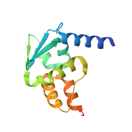 | |
UniProt & NIH Common Fund Data Resources | |||||
Find proteins for Q1H9T6 (Mus musculus) Explore Q1H9T6 Go to UniProtKB: Q1H9T6 | |||||
IMPC: MGI:2140248 | |||||
Entity Groups | |||||
| Sequence Clusters | 30% Identity50% Identity70% Identity90% Identity95% Identity100% Identity | ||||
| UniProt Group | Q1H9T6 | ||||
Sequence AnnotationsExpand | |||||
| |||||
| Length ( Å ) | Angle ( ˚ ) |
|---|---|
| a = 67.821 | α = 90 |
| b = 67.821 | β = 90 |
| c = 216.487 | γ = 120 |
| Software Name | Purpose |
|---|---|
| CNS | refinement |
| HKL-2000 | data reduction |
| HKL-2000 | data scaling |
| SOLVE | phasing |
RCSB PDB (citation) is hosted by
RCSB PDB is a member of the

