2X58

The crystal structure of MFE1 liganded with CoA


Experimental Data Snapshot

  • Method: X-RAY DIFFRACTION
  • Resolution: 2.80 Å
  • R-Value Free: 0.282 
  • R-Value Work: 0.224 
  • R-Value Observed: 0.227 

wwPDB Validation   3D Report Full Report


Ligand Structure Quality Assessment 


This is version 1.3 of the entry. See complete history


Literature

The Crystal Structure of Liganded Rat Peroxisomal Multifunctional Enzyme Type 1: A Flexible Molecule with Two Interconnected Active Sites

Kasaragod, P.Venkatesan, R.Kiema, T.R.Hiltunen, J.K.Wierenga, R.K.

(2010) J Biol Chem 285: 24089

  • DOI: https://doi.org/10.1074/jbc.M110.117606
  • Primary Citation of Related Structures:  
    2X58

  • PubMed Abstract: 

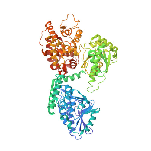
    The crystal structure of the full-length rat peroxisomal multifunctional enzyme, type 1 (rpMFE1), has been determined at 2.8 A resolution. This enzyme has three catalytic activities and two active sites. The N-terminal part has the crotonase fold, which builds the active site for the Delta(3),Delta(2)-enoyl-CoA isomerase and the Delta(2)-enoyl-CoA hydratase-1 catalytic activities, and the C-terminal part has the (3S)-hydroxyacyl-CoA dehydrogenase fold and makes the (3S)-hydroxyacyl-CoA dehydrogenase active site. rpMFE1 is a multidomain protein having five domains (A-E). The crystal structure of full-length rpMFE1 shows a flexible arrangement of the A-domain with respect to the B-E-domains. Because of a hinge region near the end of the A-domain, two different positions of the A-domain were observed for the two protein molecules (A and B) of the asymmetric unit. In the most closed conformation, the mode of binding of CoA is stabilized by domains A and B (helix-10), as seen in other crotonase fold members. Domain B, although functionally belonging to the N-terminal part, is found tightly associated with the C-terminal part, i.e. fixed to the E-domain. The two active sites of rpMFE1 are approximately 40 A apart, separated by a tunnel, characterized by an excess of positively charged side chains. Comparison of the structures of rpMFE1 with the monofunctional crotonase and (3S)-hydroxyacyl-CoA dehydrogenase superfamily enzymes, as well as with the bacterial alpha(2)beta(2)-fatty acid oxidation multienzyme complex, reveals that this tunnel could be important for substrate channeling, as observed earlier on the basis of the kinetics of rpMFE1 purified from rat liver.


  • Organizational Affiliation

    Biocenter Oulu and Department of Biochemistry, University of Oulu, FI-90014 Oulu, Finland.


Macromolecules
Find similar proteins by:  (by identity cutoff)  |  3D Structure
Entity ID: 1
MoleculeChains Sequence LengthOrganismDetailsImage
PEROXISOMAL BIFUNCTIONAL ENZYME
A, B
727Rattus norvegicusMutation(s): 0 
EC: 4.2.1.17 (PDB Primary Data), 5.3.3.8 (PDB Primary Data), 1.1.1.35 (PDB Primary Data)
UniProt
Find proteins for P07896 (Rattus norvegicus)
Explore P07896 
Go to UniProtKB:  P07896
Entity Groups  
Sequence Clusters30% Identity50% Identity70% Identity90% Identity95% Identity100% Identity
UniProt GroupP07896
Sequence Annotations
Expand
  • Reference Sequence
Small Molecules
Ligands 4 Unique
IDChains Name / Formula / InChI Key2D Diagram3D Interactions
COA
Query on COA

Download Ideal Coordinates CCD File 
D [auth A],
I [auth B]
COENZYME A
C21 H36 N7 O16 P3 S
RGJOEKWQDUBAIZ-IBOSZNHHSA-N
ADP
Query on ADP

Download Ideal Coordinates CCD File 
C [auth A]ADENOSINE-5'-DIPHOSPHATE
C10 H15 N5 O10 P2
XTWYTFMLZFPYCI-KQYNXXCUSA-N
SO4
Query on SO4

Download Ideal Coordinates CCD File 
F [auth A],
G [auth A],
H [auth A],
L [auth B],
M [auth B]
SULFATE ION
O4 S
QAOWNCQODCNURD-UHFFFAOYSA-L
GOL
Query on GOL

Download Ideal Coordinates CCD File 
E [auth A],
J [auth B],
K [auth B]
GLYCEROL
C3 H8 O3
PEDCQBHIVMGVHV-UHFFFAOYSA-N
Experimental Data & Validation

Experimental Data

  • Method: X-RAY DIFFRACTION
  • Resolution: 2.80 Å
  • R-Value Free: 0.282 
  • R-Value Work: 0.224 
  • R-Value Observed: 0.227 
  • Space Group: P 21 21 21
Unit Cell:
Length ( Å )Angle ( ˚ )
a = 65.7α = 90
b = 126.5β = 90
c = 227.07γ = 90
Software Package:
Software NamePurpose
REFMACrefinement
PHASERphasing

Structure Validation

View Full Validation Report


Ligand Structure Quality Assessment 


Entry History 

Deposition Data

Revision History  (Full details and data files)

  • Version 1.0: 2010-05-12
    Type: Initial release
  • Version 1.1: 2011-05-08
    Changes: Version format compliance
  • Version 1.2: 2011-07-13
    Changes: Version format compliance
  • Version 1.3: 2023-12-20
    Changes: Data collection, Database references, Derived calculations, Other, Refinement description