2PZI

Crystal Structure of Protein kinase PknG from Mycobacterium tuberculosis in Complex with Tetrahydrobenzothiophene AX20017


Experimental Data Snapshot

  • Method: X-RAY DIFFRACTION
  • Resolution: 2.40 Å
  • R-Value Free: 0.233 
  • R-Value Work: 0.183 
  • R-Value Observed: 0.186 

wwPDB Validation   3D Report Full Report


Ligand Structure Quality Assessment 


This is version 1.4 of the entry. See complete history


Literature

Structural basis for the specific inhibition of protein kinase G, a virulence factor of Mycobacterium tuberculosis.

Scherr, N.Honnappa, S.Kunz, G.Mueller, P.Jayachandran, R.Winkler, F.Pieters, J.Steinmetz, M.O.

(2007) Proc Natl Acad Sci U S A 104: 12151-12156

  • DOI: https://doi.org/10.1073/pnas.0702842104
  • Primary Citation of Related Structures:  
    2PZI

  • PubMed Abstract: 

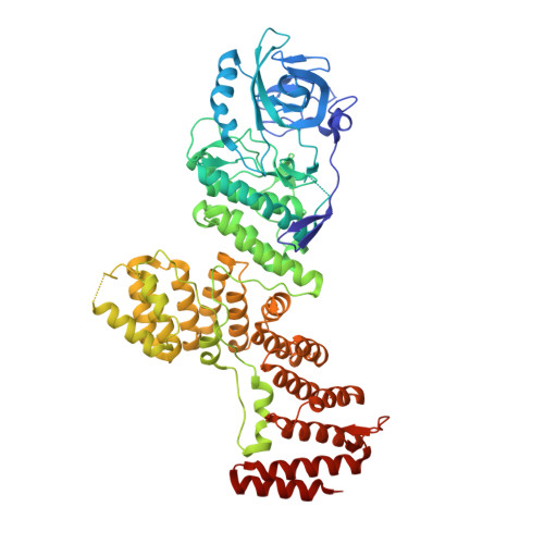
    The pathogenicity of mycobacteria such as Mycobacterium tuberculosis is closely associated with their capacity to survive within host macrophages. A crucial virulence factor for intracellular mycobacterial survival is protein kinase G (PknG), a eukaryotic-like serine/threonine protein kinase expressed by pathogenic mycobacteria that blocks the intracellular degradation of mycobacteria in lysosomes. Inhibition of PknG with the highly selective low-molecular-weight inhibitor AX20017 results in mycobacterial transfer to lysosomes and killing of the mycobacteria. Here, we report the 2.4 A x-ray crystal structure of PknG in complex with AX20017. The unique multidomain topology of PknG reveals a central kinase domain that is flanked by N- and C-terminal rubredoxin and tetratrico-peptide repeat domains, respectively. Directed mutagenesis suggests that the rubredoxin domain functions as a regulator of PknG kinase activity. The structure of PknG-AX20017 further reveals that the inhibitor is buried deep within the adenosine-binding site, targeting an active conformation of the kinase domain. Remarkably, although the topology of the kinase domain is reminiscent of eukaryotic kinases, the AX20017-binding pocket is shaped by a unique set of amino acid side chains that are not found in any human kinase. Directed mutagenesis of the unique set of residues resulted in a drastic loss of the compound's inhibitory potency. Our results explain the specific mode of action of AX20017 and demonstrate that virulence factors highly homologous to host molecules can be successfully targeted to block the proliferation of M. tuberculosis.


  • Organizational Affiliation

    Biozentrum, University of Basel, CH-4056 Basel, Switzerland.


Macromolecules
Find similar proteins by:  (by identity cutoff)  |  3D Structure
Entity ID: 1
MoleculeChains Sequence LengthOrganismDetailsImage
Probable serine/threonine-protein kinase pknG
A, B
681Mycobacterium tuberculosisMutation(s): 0 
Gene Names: pknG
EC: 2.7.11.1
UniProt
Find proteins for P9WI73 (Mycobacterium tuberculosis (strain ATCC 25618 / H37Rv))
Explore P9WI73 
Go to UniProtKB:  P9WI73
Entity Groups  
Sequence Clusters30% Identity50% Identity70% Identity90% Identity95% Identity100% Identity
UniProt GroupP9WI73
Sequence Annotations
Expand
  • Reference Sequence
Small Molecules
Ligands 4 Unique
IDChains Name / Formula / InChI Key2D Diagram3D Interactions
AXX
Query on AXX

Download Ideal Coordinates CCD File 
E [auth A],
K [auth B]
2-[(CYCLOPROPYLCARBONYL)AMINO]-4,5,6,7-TETRAHYDRO-1-BENZOTHIOPHENE-3-CARBOXAMIDE
C13 H16 N2 O2 S
VATFNEMGBRWLHI-UHFFFAOYSA-N
CD
Query on CD

Download Ideal Coordinates CCD File 
D [auth A],
J [auth B]
CADMIUM ION
Cd
WLZRMCYVCSSEQC-UHFFFAOYSA-N
GOL
Query on GOL

Download Ideal Coordinates CCD File 
F [auth A]
G [auth A]
H [auth A]
I [auth A]
L [auth B]
F [auth A],
G [auth A],
H [auth A],
I [auth A],
L [auth B],
M [auth B]
GLYCEROL
C3 H8 O3
PEDCQBHIVMGVHV-UHFFFAOYSA-N
CL
Query on CL

Download Ideal Coordinates CCD File 
C [auth A]CHLORIDE ION
Cl
VEXZGXHMUGYJMC-UHFFFAOYSA-M
Binding Affinity Annotations 
IDSourceBinding Affinity
AXX Binding MOAD:  2PZI IC50: 3000 (nM) from 1 assay(s)
PDBBind:  2PZI IC50: 800 (nM) from 1 assay(s)
Experimental Data & Validation

Experimental Data

  • Method: X-RAY DIFFRACTION
  • Resolution: 2.40 Å
  • R-Value Free: 0.233 
  • R-Value Work: 0.183 
  • R-Value Observed: 0.186 
  • Space Group: P 65
Unit Cell:
Length ( Å )Angle ( ˚ )
a = 122.553α = 90
b = 122.553β = 90
c = 243.749γ = 120
Software Package:
Software NamePurpose
REFMACrefinement
MAR345data collection
XDSdata reduction
XSCALEdata scaling
SHARPphasing

Structure Validation

View Full Validation Report


Ligand Structure Quality Assessment 


Entry History 

Deposition Data

Revision History  (Full details and data files)

  • Version 1.0: 2007-07-24
    Type: Initial release
  • Version 1.1: 2008-05-01
    Changes: Version format compliance
  • Version 1.2: 2011-07-13
    Changes: Advisory, Version format compliance
  • Version 1.3: 2017-10-18
    Changes: Refinement description
  • Version 1.4: 2024-02-21
    Changes: Data collection, Database references, Derived calculations