2OVR

Structure of the Skp1-Fbw7-CyclinEdegN complex


Experimental Data Snapshot

  • Method: X-RAY DIFFRACTION
  • Resolution: 2.50 Å
  • R-Value Free: 0.251 
  • R-Value Work: 0.225 
  • R-Value Observed: 0.225 

wwPDB Validation   3D Report Full Report


This is version 1.4 of the entry. See complete history


Literature

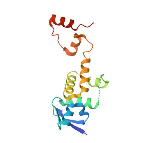
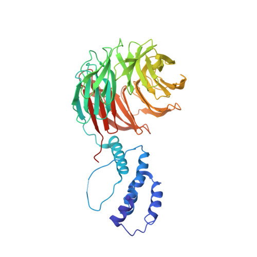
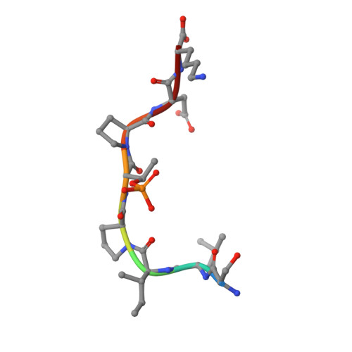
Structure of a Fbw7-Skp1-Cyclin E Complex: Multisite-Phosphorylated Substrate Recognition by SCF Ubiquitin Ligases

Hao, B.Oehlmann, S.Sowa, M.E.Harper, J.W.Pavletich, N.P.

(2007) Mol Cell 26: 131-143

  • DOI: https://doi.org/10.1016/j.molcel.2007.02.022
  • Primary Citation of Related Structures:  
    2OVP, 2OVQ, 2OVR

  • PubMed Abstract: 

    The ubiquitin-mediated proteolysis of cyclin E plays a central role in cell-cycle progression, and cyclin E accumulation is a common event in cancer. Cyclin E degradation is triggered by multisite phosphorylation, which induces binding to the SCF(Fbw7) ubiquitin ligase complex. Structures of the Skp1-Fbw7 complex bound to cyclin E peptides identify a doubly phosphorylated pThr380/pSer384 cyclin E motif as an optimal, high-affinity degron and a singly phosphorylated pThr62 motif as a low-affinity one. Biochemical data indicate that the closely related yeast SCF(Cdc4) complex recognizes the multisite phosphorylated Sic1 substrate similarly and identify three doubly phosphorylated Sic1 degrons, each capable of high-affinity interactions with two Cdc4 phosphate binding sites. A model that explains the role of multiple cyclin E/Sic1 degrons is provided by the findings that Fbw7 and Cdc4 dimerize, that Fbw7 dimerization enhances the turnover of a weakly associated cyclin E in vivo, and that Cdc4 dimerization increases the rate and processivity of Sic1 ubiquitination in vitro.


  • Organizational Affiliation

    Structural Biology Program, Memorial Sloan-Kettering Cancer Center, New York, NY 10021, USA.


Macromolecules
Find similar proteins by:  (by identity cutoff)  |  3D Structure
Entity ID: 1
MoleculeChains Sequence LengthOrganismDetailsImage
S-phase kinase-associated protein 1A149Homo sapiensMutation(s): 0 
Gene Names: SKP1AEMC19OCP2SKP1TCEB1L
UniProt & NIH Common Fund Data Resources
Find proteins for P63208 (Homo sapiens)
Explore P63208 
Go to UniProtKB:  P63208
PHAROS:  P63208
GTEx:  ENSG00000113558 
Entity Groups  
Sequence Clusters30% Identity50% Identity70% Identity90% Identity95% Identity100% Identity
UniProt GroupP63208
Sequence Annotations
Expand
  • Reference Sequence
Find similar proteins by:  (by identity cutoff)  |  3D Structure
Entity ID: 2
MoleculeChains Sequence LengthOrganismDetailsImage
F-box/WD repeat protein 7445Homo sapiensMutation(s): 0 
Gene Names: FBXW7FBW7FBX30SEL10
UniProt & NIH Common Fund Data Resources
Find proteins for Q969H0 (Homo sapiens)
Explore Q969H0 
Go to UniProtKB:  Q969H0
PHAROS:  Q969H0
GTEx:  ENSG00000109670 
Entity Groups  
Sequence Clusters30% Identity50% Identity70% Identity90% Identity95% Identity100% Identity
UniProt GroupQ969H0
Sequence Annotations
Expand
  • Reference Sequence

Find similar proteins by:  Sequence   |   3D Structure  

Entity ID: 3
MoleculeChains Sequence LengthOrganismDetailsImage
cyclinE N-terminal degron8N/AMutation(s): 0 
Sequence Annotations
Expand
  • Reference Sequence
Small Molecules
Ligands 1 Unique
IDChains Name / Formula / InChI Key2D Diagram3D Interactions
SO4
Query on SO4

Download Ideal Coordinates CCD File 
D [auth B]
E [auth B]
F [auth B]
G [auth B]
H [auth B]
D [auth B],
E [auth B],
F [auth B],
G [auth B],
H [auth B],
I [auth B],
J [auth B],
K [auth B],
L [auth B],
M [auth B],
N [auth B],
O [auth B]
SULFATE ION
O4 S
QAOWNCQODCNURD-UHFFFAOYSA-L
Modified Residues  1 Unique
IDChains TypeFormula2D DiagramParent
TPO
Query on TPO
C
L-PEPTIDE LINKINGC4 H10 N O6 PTHR
Experimental Data & Validation

Experimental Data

  • Method: X-RAY DIFFRACTION
  • Resolution: 2.50 Å
  • R-Value Free: 0.251 
  • R-Value Work: 0.225 
  • R-Value Observed: 0.225 
  • Space Group: I 41 2 2
Unit Cell:
Length ( Å )Angle ( ˚ )
a = 232.558α = 90
b = 232.558β = 90
c = 107.659γ = 90
Software Package:
Software NamePurpose
ADSCdata collection
AMoREphasing
CNSrefinement
DENZOdata reduction
SCALEPACKdata scaling

Structure Validation

View Full Validation Report


Entry History 

Deposition Data

Revision History  (Full details and data files)

  • Version 1.0: 2007-04-24
    Type: Initial release
  • Version 1.1: 2008-05-01
    Changes: Version format compliance
  • Version 1.2: 2011-07-13
    Changes: Version format compliance
  • Version 1.3: 2011-11-16
    Changes: Atomic model
  • Version 1.4: 2024-04-03
    Changes: Data collection, Database references, Derived calculations, Refinement description