2OO9

crystal structure of the UBA domain from human c-Cbl ubiquitin ligase


Experimental Data Snapshot

  • Method: X-RAY DIFFRACTION
  • Resolution: 2.10 Å
  • R-Value Free: 0.260 
  • R-Value Work: 0.216 
  • R-Value Observed: 0.218 

wwPDB Validation   3D Report Full Report


This is version 1.3 of the entry. See complete history


Literature

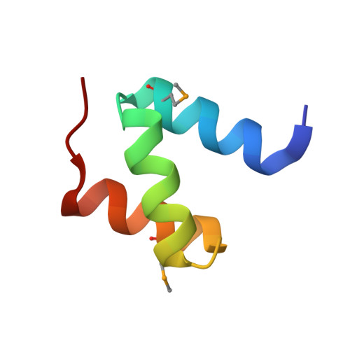
Structural basis for UBA-mediated dimerization of c-Cbl ubiquitin ligase.

Kozlov, G.Peschard, P.Zimmerman, B.Lin, T.Moldoveanu, T.Mansur-Azzam, N.Gehring, K.Park, M.

(2007) J Biol Chem 282: 27547-27555

  • DOI: https://doi.org/10.1074/jbc.M703333200
  • Primary Citation of Related Structures:  
    2OO9

  • PubMed Abstract: 

    Ligand-induced down-regulation by the ubiquitin-protein ligases, c-Cbl and Cbl-b, controls signaling downstream from many receptor-tyrosine kinases (RTK). Cbl proteins bind to phosphotyrosine residues on activated RTKs to affect ligand-dependent ubiquitylation of these receptors targeting them for degradation in the lysosome. Both c-Cbl and Cbl-b contain a ubiquitin-associated (UBA) domain, which is important for Cbl dimerization and tyrosine phosphorylation; however, the mechanism of UBA-mediated dimerization and its requirement for Cbl biological activity is unclear. Here, we report the crystal structure of the UBA domain of c-Cbl refined to 2.1-A resolution. The structure reveals the protein is a symmetric dimer tightly packed along a large hydrophobic surface formed by helices 2 and 3. NMR chemical shift mapping reveals heterodimerization can occur with the related Cbl-b UBA domain via the same surface employed for homodimerization. Disruption of c-Cbl dimerization by site-directed mutagenesis impairs c-Cbl phosphorylation following activation of the Met/hepatocyte growth factor RTK and c-Cbl-dependent ubiquitination of Met. This provides direct evidence for a role of Cbl dimerization in terminating signaling following activation of RTKs.


  • Organizational Affiliation

    Department of Biochemistry, McGill University.


Macromolecules
Find similar proteins by:  (by identity cutoff)  |  3D Structure
Entity ID: 1
MoleculeChains Sequence LengthOrganismDetailsImage
E3 ubiquitin-protein ligase CBL
A, B, C
46Homo sapiensMutation(s): 2 
Gene Names: CBLCBL2RNF55
EC: 6.3.2
UniProt & NIH Common Fund Data Resources
Find proteins for P22681 (Homo sapiens)
Explore P22681 
Go to UniProtKB:  P22681
PHAROS:  P22681
GTEx:  ENSG00000110395 
Entity Groups  
Sequence Clusters30% Identity50% Identity70% Identity90% Identity95% Identity100% Identity
UniProt GroupP22681
Sequence Annotations
Expand
  • Reference Sequence
Small Molecules
Modified Residues  1 Unique
IDChains TypeFormula2D DiagramParent
MSE
Query on MSE
A, B, C
L-PEPTIDE LINKINGC5 H11 N O2 SeMET
Experimental Data & Validation

Experimental Data

  • Method: X-RAY DIFFRACTION
  • Resolution: 2.10 Å
  • R-Value Free: 0.260 
  • R-Value Work: 0.216 
  • R-Value Observed: 0.218 
  • Space Group: P 41 21 2
Unit Cell:
Length ( Å )Angle ( ˚ )
a = 82.025α = 90
b = 82.025β = 90
c = 56.194γ = 90
Software Package:
Software NamePurpose
REFMACrefinement
HKL-2000data collection
HKL-2000data reduction
HKL-2000data scaling
SOLVEphasing

Structure Validation

View Full Validation Report


Entry History 

Deposition Data

Revision History  (Full details and data files)

  • Version 1.0: 2007-02-06
    Type: Initial release
  • Version 1.1: 2008-05-01
    Changes: Version format compliance
  • Version 1.2: 2011-07-13
    Changes: Advisory, Version format compliance
  • Version 1.3: 2023-12-27
    Changes: Data collection, Database references, Derived calculations