2LB3

Structure of the WW domain of PIN1 in complex with a human phosphorylated Smad3 derived peptide


Experimental Data Snapshot

  • Method: SOLUTION NMR
  • Conformers Calculated: 300 
  • Conformers Submitted: 20 
  • Selection Criteria: structures with acceptable covalent geometry 

wwPDB Validation   3D Report Full Report


This is version 1.1 of the entry. See complete history


Literature

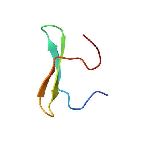
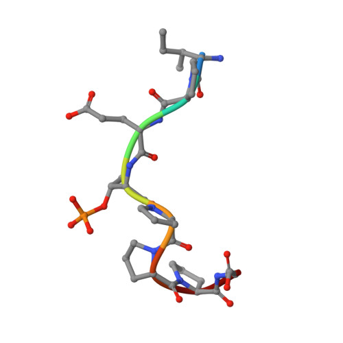
A Smad action turnover switch operated by WW domain readers of a phosphoserine code.

Aragon, E.Goerner, N.Zaromytidou, A.I.Xi, Q.Escobedo, A.Massague, J.Macias, M.J.

(2011) Genes Dev 25: 1275-1288

  • DOI: https://doi.org/10.1101/gad.2060811
  • Primary Citation of Related Structures:  
    2LAJ, 2LAW, 2LAX, 2LAY, 2LAZ, 2LB0, 2LB1, 2LB2, 2LB3

  • PubMed Abstract: 

    When directed to the nucleus by TGF-β or BMP signals, Smad proteins undergo cyclin-dependent kinase 8/9 (CDK8/9) and glycogen synthase kinase-3 (GSK3) phosphorylations that mediate the binding of YAP and Pin1 for transcriptional action, and of ubiquitin ligases Smurf1 and Nedd4L for Smad destruction. Here we demonstrate that there is an order of events-Smad activation first and destruction later-and that it is controlled by a switch in the recognition of Smad phosphoserines by WW domains in their binding partners. In the BMP pathway, Smad1 phosphorylation by CDK8/9 creates binding sites for the WW domains of YAP, and subsequent phosphorylation by GSK3 switches off YAP binding and adds binding sites for Smurf1 WW domains. Similarly, in the TGF-β pathway, Smad3 phosphorylation by CDK8/9 creates binding sites for Pin1 and GSK3, then adds sites to enhance Nedd4L binding. Thus, a Smad phosphoserine code and a set of WW domain code readers provide an efficient solution to the problem of coupling TGF-β signal delivery to turnover of the Smad signal transducers.


  • Organizational Affiliation

    Structural and Computational Biology Programme, Institute for Research in Biomedicine, Barcelona, Spain.


Macromolecules
Find similar proteins by:  (by identity cutoff)  |  3D Structure
Entity ID: 1
MoleculeChains Sequence LengthOrganismDetailsImage
Peptidyl-prolyl cis-trans isomerase NIMA-interacting 136Homo sapiensMutation(s): 0 
Gene Names: PIN1
EC: 5.2.1.8
UniProt & NIH Common Fund Data Resources
Find proteins for Q13526 (Homo sapiens)
Explore Q13526 
Go to UniProtKB:  Q13526
PHAROS:  Q13526
GTEx:  ENSG00000127445 
Entity Groups  
Sequence Clusters30% Identity50% Identity70% Identity90% Identity95% Identity100% Identity
UniProt GroupQ13526
Sequence Annotations
Expand
  • Reference Sequence

Find similar proteins by:  Sequence   |   3D Structure  

Entity ID: 2
MoleculeChains Sequence LengthOrganismDetailsImage
Mothers against decapentaplegic homolog 28Homo sapiensMutation(s): 0 
UniProt & NIH Common Fund Data Resources
Find proteins for Q15796 (Homo sapiens)
Explore Q15796 
Go to UniProtKB:  Q15796
PHAROS:  Q15796
GTEx:  ENSG00000175387 
Entity Groups  
UniProt GroupQ15796
Sequence Annotations
Expand
  • Reference Sequence
Small Molecules
Modified Residues  1 Unique
IDChains TypeFormula2D DiagramParent
TPO
Query on TPO
B
L-PEPTIDE LINKINGC4 H10 N O6 PTHR
Experimental Data & Validation

Experimental Data

  • Method: SOLUTION NMR
  • Conformers Calculated: 300 
  • Conformers Submitted: 20 
  • Selection Criteria: structures with acceptable covalent geometry 

Structure Validation

View Full Validation Report


Entry History 

Deposition Data

Revision History  (Full details and data files)

  • Version 1.0: 2011-07-06
    Type: Initial release
  • Version 1.1: 2011-07-13
    Changes: Version format compliance