2E52

Crystal structural analysis of HindIII restriction endonuclease in complex with cognate DNA at 2.0 angstrom resolution


Experimental Data Snapshot

  • Method: X-RAY DIFFRACTION
  • Resolution: 2.00 Å
  • R-Value Free: 0.217 
  • R-Value Work: 0.175 
  • R-Value Observed: 0.177 

wwPDB Validation   3D Report Full Report


This is version 1.3 of the entry. See complete history


Literature

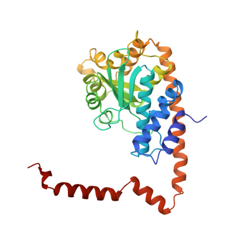
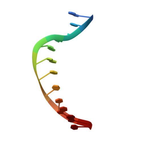
Structures of restriction endonuclease HindIII in complex with its cognate DNA and divalent cations

Watanabe, N.Takasaki, Y.Sato, C.Ando, S.Tanaka, I.

(2009) Acta Crystallogr D Biol Crystallogr 65: 1326-1333

  • DOI: https://doi.org/10.1107/S0907444909041134
  • Primary Citation of Related Structures:  
    2E52, 3A4K

  • PubMed Abstract: 

    The three-dimensional crystal structures of HindIII bound to its cognate DNA with and without divalent cations were solved at 2.17 and 2.00 A resolution, respectively. HindIII forms a dimer. The structures showed that HindIII belongs to the EcoRI-like (alpha-class) subfamily of type II restriction endonucleases. The cognate DNA-complex structures revealed the specific DNA-recognition mechanism of HindIII by which it recognizes the palindromic sequence A/AGCTT. In the Mg(2+) ion-soaked structure the DNA was cleaved and two ions were bound at each active site, corresponding to the two-metal-ion mechanism.


  • Organizational Affiliation

    Department of Biotechnology and Biomaterial Chemistry, Graduate School of Engineering, Nagoya University, Japan. nobuhisa@nagoya-u.jp


Macromolecules

Find similar proteins by:  (by identity cutoff)  |  3D Structure
Entity ID: 1
MoleculeChains Sequence LengthOrganismDetailsImage
Type II restriction enzyme HindIIIA,
B [auth C],
C [auth B],
D
300Haemophilus influenzae Rd KW20Mutation(s): 0 
Gene Names: hindIIIR
EC: 3.1.21.4
UniProt
Find proteins for P43870 (Haemophilus influenzae (strain ATCC 51907 / DSM 11121 / KW20 / Rd))
Explore P43870 
Go to UniProtKB:  P43870
Entity Groups  
Sequence Clusters30% Identity50% Identity70% Identity90% Identity95% Identity100% Identity
UniProt GroupP43870
Sequence Annotations
Expand
  • Reference Sequence

Find similar nucleic acids by:  Sequence   |   3D Structure  

Entity ID: 2
MoleculeChains LengthOrganismImage
DNA (5'-D(*DGP*DCP*DCP*DAP*DAP*DGP*DCP*DTP*DTP*DGP*DGP*DC)-3')
E, F, G, H, I
E, F, G, H, I, J
12N/A
Sequence Annotations
Expand
  • Reference Sequence
Experimental Data & Validation

Experimental Data

  • Method: X-RAY DIFFRACTION
  • Resolution: 2.00 Å
  • R-Value Free: 0.217 
  • R-Value Work: 0.175 
  • R-Value Observed: 0.177 
  • Space Group: P 1 21 1
Unit Cell:
Length ( Å )Angle ( ˚ )
a = 83.125α = 90
b = 131.532β = 111.2
c = 93.718γ = 90
Software Package:
Software NamePurpose
HKL-2000data collection
SOLVEphasing
REFMACrefinement
HKL-2000data reduction
HKL-2000data scaling

Structure Validation

View Full Validation Report


Entry History 

Deposition Data

Revision History  (Full details and data files)

  • Version 1.0: 2007-12-18
    Type: Initial release
  • Version 1.1: 2011-07-13
    Changes: Source and taxonomy, Version format compliance
  • Version 1.2: 2014-03-05
    Changes: Database references
  • Version 1.3: 2024-03-13
    Changes: Data collection, Database references, Derived calculations