2DYT

The crystal structure of Saccharomyces cerevisiae Atg3


Experimental Data Snapshot

  • Method: X-RAY DIFFRACTION
  • Resolution: 2.50 Å
  • R-Value Free: 0.244 
  • R-Value Work: 0.207 
  • R-Value Observed: 0.207 

wwPDB Validation   3D Report Full Report


This is version 1.3 of the entry. See complete history


Literature

The crystal structure of Atg3, an autophagy-related ubiquitin carrier protein (E2) enzyme that mediates Atg8 lipidation

Yamada, Y.Suzuki, N.N.Hanada, T.Ichimura, Y.Kumeta, H.Fujioka, Y.Ohsumi, Y.Inagaki, F.

(2007) J Biol Chem 282: 8036-8043

  • DOI: https://doi.org/10.1074/jbc.M611473200
  • Primary Citation of Related Structures:  
    2DYT

  • PubMed Abstract: 

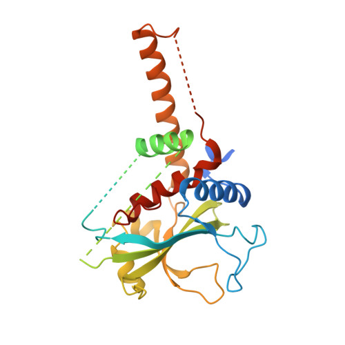
    Atg3 is an E2-like enzyme that catalyzes the conjugation of Atg8 and phosphatidylethanolamine (PE). The Atg8-PE conjugate is essential for autophagy, which is the bulk degradation process of cytoplasmic components by the vacuolar/lysosomal system. We report here the crystal structure of Saccharomyces cerevisiae Atg3 at 2.5-A resolution. Atg3 has an alpha/beta-fold, and its core region is topologically similar to canonical E2 enzymes. Atg3 has two regions inserted in the core region, one of which consists of approximately 80 residues and has a random coil structure in solution and another with a long alpha-helical structure that protrudes from the core region as far as 30 A. In vivo and in vitro analyses suggested that the former region is responsible for binding Atg7, an E1-like enzyme, and that the latter is responsible for binding Atg8. A sulfate ion was bound near the catalytic cysteine of Atg3, suggesting a possible binding site for the phosphate moiety of PE. The structure of Atg3 provides a molecular basis for understanding the unique lipidation reaction that Atg3 carries out.


  • Organizational Affiliation

    Department of Structural Biology, Graduate School of Pharmaceutical Sciences, Hokkaido University, N-12, W-6, Kita-ku, Sapporo 060-0812, Japan.


Macromolecules
Find similar proteins by:  (by identity cutoff)  |  3D Structure
Entity ID: 1
MoleculeChains Sequence LengthOrganismDetailsImage
Autophagy-related protein 3312Saccharomyces cerevisiaeMutation(s): 0 
UniProt
Find proteins for P40344 (Saccharomyces cerevisiae (strain ATCC 204508 / S288c))
Explore P40344 
Go to UniProtKB:  P40344
Entity Groups  
Sequence Clusters30% Identity50% Identity70% Identity90% Identity95% Identity100% Identity
UniProt GroupP40344
Sequence Annotations
Expand
  • Reference Sequence
Experimental Data & Validation

Experimental Data

  • Method: X-RAY DIFFRACTION
  • Resolution: 2.50 Å
  • R-Value Free: 0.244 
  • R-Value Work: 0.207 
  • R-Value Observed: 0.207 
  • Space Group: P 41
Unit Cell:
Length ( Å )Angle ( ˚ )
a = 59.116α = 90
b = 59.116β = 90
c = 115.316γ = 90
Software Package:
Software NamePurpose
CNSrefinement
HKL-2000data collection
HKL-2000data reduction
HKL-2000data scaling
SOLVEphasing

Structure Validation

View Full Validation Report


Entry History 

Deposition Data

Revision History  (Full details and data files)

  • Version 1.0: 2007-01-23
    Type: Initial release
  • Version 1.1: 2007-10-08
    Changes: Version format compliance
  • Version 1.2: 2011-07-13
    Changes: Version format compliance
  • Version 1.3: 2024-03-13
    Changes: Data collection, Database references, Derived calculations