2BRQ

Crystal structure of the filamin A repeat 21 complexed with the integrin beta7 cytoplasmic tail peptide


Experimental Data Snapshot

  • Method: X-RAY DIFFRACTION
  • Resolution: 2.10 Å
  • R-Value Free: 0.243 
  • R-Value Work: 0.192 
  • R-Value Observed: 0.197 

wwPDB Validation   3D Report Full Report


Ligand Structure Quality Assessment 


This is version 1.3 of the entry. See complete history


Literature

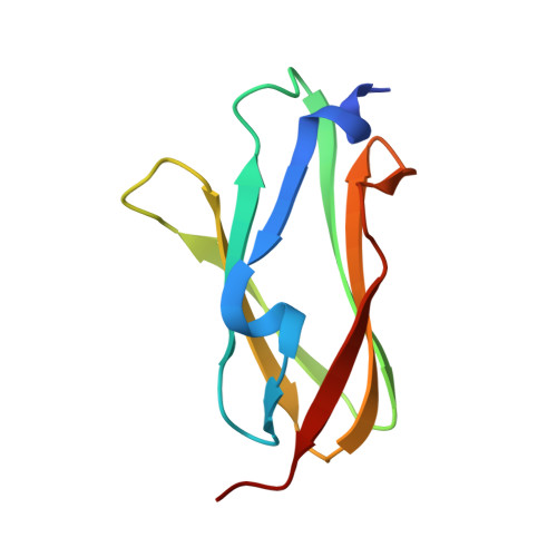
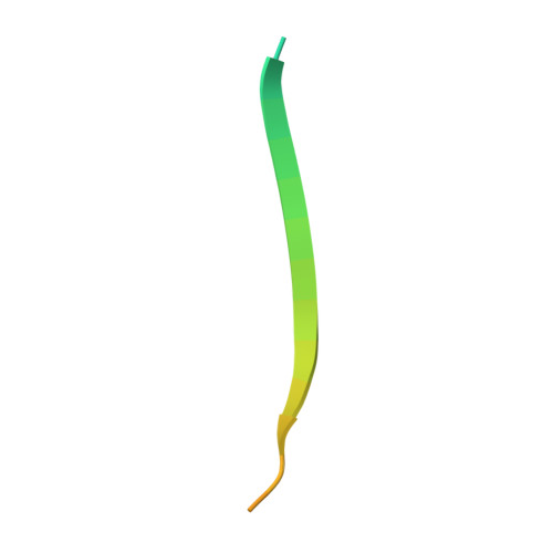
The Molecular Basis of Filamin Binding to Integrins and Competition with Talin.

Kiema, T.Lad, Y.Jiang, P.Oxley, C.L.Baldassarre, M.Wegener, K.L.Campbell, I.D.Ylanne, J.Calderwood, D.A.

(2006) Mol Cell 21: 337

  • DOI: https://doi.org/10.1016/j.molcel.2006.01.011
  • Primary Citation of Related Structures:  
    2BRQ

  • PubMed Abstract: 

    The ability of adhesion receptors to transmit biochemical signals and mechanical force across cell membranes depends on interactions with the actin cytoskeleton. Filamins are large, actin-crosslinking proteins that connect multiple transmembrane and signaling proteins to the cytoskeleton. Here, we describe the high-resolution structure of an interface between filamin A and an integrin adhesion receptor. When bound, the integrin beta cytoplasmic tail forms an extended beta strand that interacts with beta strands C and D of the filamin immunoglobulin-like domain (IgFLN) 21. This interface is common to many integrins, and we suggest it is a prototype for other IgFLN domain interactions. Notably, the structurally defined filamin binding site overlaps with that of the integrin-regulator talin, and these proteins compete for binding to integrin tails, allowing integrin-filamin interactions to impact talin-dependent integrin activation. Phosphothreonine-mimicking mutations inhibit filamin, but not talin, binding, indicating that kinases may modulate this competition and provide additional means to control integrin functions.


  • Organizational Affiliation

    Biocenter Oulu, Department of Biochemistry, University of Oulu, FIN-90014 Oulu, Finland.


Macromolecules
Find similar proteins by:  (by identity cutoff)  |  3D Structure
Entity ID: 1
MoleculeChains Sequence LengthOrganismDetailsImage
FILAMIN A
A, B
97Homo sapiensMutation(s): 0 
UniProt & NIH Common Fund Data Resources
Find proteins for P21333 (Homo sapiens)
Explore P21333 
Go to UniProtKB:  P21333
PHAROS:  P21333
GTEx:  ENSG00000196924 
Entity Groups  
Sequence Clusters30% Identity50% Identity70% Identity90% Identity95% Identity100% Identity
UniProt GroupP21333
Sequence Annotations
Expand
  • Reference Sequence
Find similar proteins by:  (by identity cutoff)  |  3D Structure
Entity ID: 2
MoleculeChains Sequence LengthOrganismDetailsImage
INTEGRIN BETA-7 SUBUNIT
C, D
31Homo sapiensMutation(s): 0 
UniProt & NIH Common Fund Data Resources
Find proteins for P26010 (Homo sapiens)
Explore P26010 
Go to UniProtKB:  P26010
PHAROS:  P26010
GTEx:  ENSG00000139626 
Entity Groups  
Sequence Clusters30% Identity50% Identity70% Identity90% Identity95% Identity100% Identity
UniProt GroupP26010
Sequence Annotations
Expand
  • Reference Sequence
Experimental Data & Validation

Experimental Data

  • Method: X-RAY DIFFRACTION
  • Resolution: 2.10 Å
  • R-Value Free: 0.243 
  • R-Value Work: 0.192 
  • R-Value Observed: 0.197 
  • Space Group: P 1 21 1
Unit Cell:
Length ( Å )Angle ( ˚ )
a = 41.97α = 90
b = 60β = 110.17
c = 51.57γ = 90
Software Package:
Software NamePurpose
REFMACrefinement
XDSdata reduction
PHASERphasing

Structure Validation

View Full Validation Report


Ligand Structure Quality Assessment 


Entry History 

Deposition Data

Revision History  (Full details and data files)

  • Version 1.0: 2006-02-07
    Type: Initial release
  • Version 1.1: 2011-07-13
    Changes: Advisory, Version format compliance
  • Version 1.2: 2012-02-29
    Changes: Other
  • Version 1.3: 2023-12-13
    Changes: Data collection, Database references, Derived calculations, Other, Refinement description