An electrical potential in the access channel of catalases enhances catalysis
Chelikani, P., Carpena, X., Fita, I., Loewen, P.C.(2003) J Biol Chem 278: 31290-31296
- PubMed: 12777389
- DOI: https://doi.org/10.1074/jbc.M304076200
- Primary Citation of Related Structures:
1P7Y, 1P7Z, 1P80, 1P81, 1QWS - PubMed Abstract:
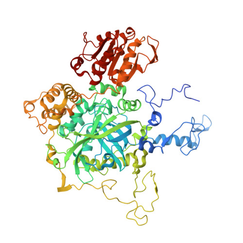
Substrate H2O2 must gain access to the deeply buried active site of catalases through channels of 30-50 A in length. The most prominent or main channel approaches the active site perpendicular to the plane of the heme and contains a number of residues that are conserved in all catalases. Changes in Val169, 8 A from the heme in catalase HPII from Escherichia coli, introducing smaller, larger or polar side chains reduces the catalase activity. Changes in Asp181, 12 A from the heme, reduces activity by up to 90% if the negatively charged side chain is removed when Ala, Gln, Ser, Asn, or Ile are the substituted residues. Only the D181E variant retains wild type activity. Determination of the crystal structures of the Glu181, Ala181, Ser181, and Gln181 variants of HPII reveals lower water occupancy in the main channel of the less active variants, particularly at the position forming the sixth ligand to the heme iron and in the hydrophobic, constricted region adjacent to Val169. It is proposed that an electrical potential exists between the negatively charged aspartate (or glutamate) side chain at position 181 and the positively charged heme iron 12 A distant. The potential field acts upon the electrical dipoles of water generating a common orientation that favors hydrogen bond formation and promotes interaction with the heme iron. Substrate hydrogen peroxide would be affected similarly and would enter the active site oriented optimally for interaction with active site residues.
Organizational Affiliation:
Department of Microbiology, University of Manitoba, Winnipeg, Manitoba R3T 2N2, Canada.