1L9L

GRANULYSIN FROM HUMAN CYTOLYTIC T LYMPHOCYTES


Experimental Data Snapshot

  • Method: X-RAY DIFFRACTION
  • Resolution: 0.92 Å
  • R-Value Free: 0.190 
  • R-Value Work: 0.137 
  • R-Value Observed: 0.138 

wwPDB Validation   3D Report Full Report


Ligand Structure Quality Assessment 


This is version 1.3 of the entry. See complete history


Literature

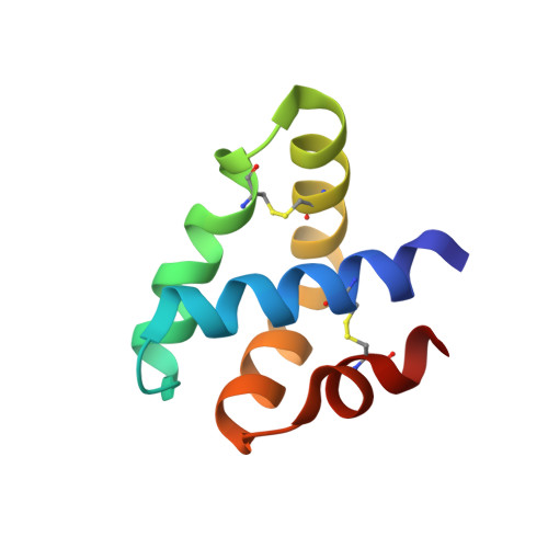
Granulysin Crystal Structure and a Structure-Derived Lytic Mechanism

Anderson, D.H.Sawaya, M.R.Cascio, D.Ernst, W.Modlin, R.Krensky, A.Eisenberg, D.

(2002) J Mol Biol 325: 355-365

  • DOI: https://doi.org/10.1016/s0022-2836(02)01234-2
  • Primary Citation of Related Structures:  
    1L9L

  • PubMed Abstract: 

    Our crystal structure of granulysin suggests a mechanism for lysis of bacterial membranes by granulysin, a 74-residue basic protein from human cytolytic T lymphocyte and natural killer cells. We determined the initial crystal structure of selenomethionyl granulysin by MAD phasing at 2A resolution. We present the structure model refined using native diffraction data to 0.96A resolution. The five-helical bundle of granulysin resembles other "saposin folds" (such as NK-lysin). Positive charges distribute in a ring around the granulysin molecule, and one face has net positive charge. Sulfate ions bind near the segment of the molecule identified as most membrane-lytic and of highest hydrophobic moment. The ion locations may indicate granulysin's orientation of initial approach towards the membrane. The crystal packing reveals one way to pack a sheet of granulysin molecules at the cell surface for a concerted lysis effort. The energy of binding granulysin charges to the bacterial membrane could drive the subsequent lytic processes. The loosely packed core facilitates a hinge or scissors motion towards exposure of hydrophobic surface that we propose tunnels the granulysin into the fracturing target membrane.


  • Organizational Affiliation

    Howard Hughes Medical Institute, 5-748 MacDonald, Box 951662, Los Angeles, CA 90095-1662, USA. dha@mbi.ucla.edu


Macromolecules
Find similar proteins by:  (by identity cutoff)  |  3D Structure
Entity ID: 1
MoleculeChains Sequence LengthOrganismDetailsImage
Granulysin74Homo sapiensMutation(s): 0 
UniProt & NIH Common Fund Data Resources
Find proteins for P22749 (Homo sapiens)
Explore P22749 
Go to UniProtKB:  P22749
PHAROS:  P22749
GTEx:  ENSG00000115523 
Entity Groups  
Sequence Clusters30% Identity50% Identity70% Identity90% Identity95% Identity100% Identity
UniProt GroupP22749
Sequence Annotations
Expand
  • Reference Sequence
Small Molecules
Ligands 3 Unique
IDChains Name / Formula / InChI Key2D Diagram3D Interactions
MPO
Query on MPO

Download Ideal Coordinates CCD File 
F [auth A]3[N-MORPHOLINO]PROPANE SULFONIC ACID
C7 H15 N O4 S
DVLFYONBTKHTER-UHFFFAOYSA-N
SO4
Query on SO4

Download Ideal Coordinates CCD File 
B [auth A],
C [auth A],
D [auth A],
E [auth A]
SULFATE ION
O4 S
QAOWNCQODCNURD-UHFFFAOYSA-L
EOH
Query on EOH

Download Ideal Coordinates CCD File 
G [auth A],
H [auth A],
I [auth A]
ETHANOL
C2 H6 O
LFQSCWFLJHTTHZ-UHFFFAOYSA-N
Experimental Data & Validation

Experimental Data

  • Method: X-RAY DIFFRACTION
  • Resolution: 0.92 Å
  • R-Value Free: 0.190 
  • R-Value Work: 0.137 
  • R-Value Observed: 0.138 
  • Space Group: P 1 21 1
Unit Cell:
Length ( Å )Angle ( ˚ )
a = 23.265α = 90
b = 60.436β = 90.99
c = 23.591γ = 90
Software Package:
Software NamePurpose
DENZOdata reduction
SCALEPACKdata scaling
SHELXDphasing
MLPHAREphasing
DMmodel building
ARP/wARPmodel building
SHELXL-97refinement
DMphasing

Structure Validation

View Full Validation Report


Ligand Structure Quality Assessment 


Entry History 

Deposition Data

Revision History  (Full details and data files)

  • Version 1.0: 2002-11-06
    Type: Initial release
  • Version 1.1: 2008-04-28
    Changes: Version format compliance
  • Version 1.2: 2011-07-13
    Changes: Version format compliance
  • Version 1.3: 2017-09-13
    Changes: Refinement description