1Y57

Structure of unphosphorylated c-Src in complex with an inhibitor


Experimental Data Snapshot

  • Method: X-RAY DIFFRACTION
  • Resolution: 1.91 Å
  • R-Value Free: 0.213 
  • R-Value Work: 0.188 
  • R-Value Observed: 0.189 

wwPDB Validation   3D Report Full Report


Ligand Structure Quality Assessment 


This is version 1.4 of the entry. See complete history


Literature

The Crystal Structure of a c-Src Complex in an Active Conformation Suggests Possible Steps in c-Src Activation

Cowan-Jacob, S.W.Fendrich, G.Manley, P.W.Jahnke, W.Fabbro, D.Liebetanz, J.Meyer, T.

(2005) Structure 13: 861-871

  • DOI: https://doi.org/10.1016/j.str.2005.03.012
  • Primary Citation of Related Structures:  
    1Y57

  • PubMed Abstract: 

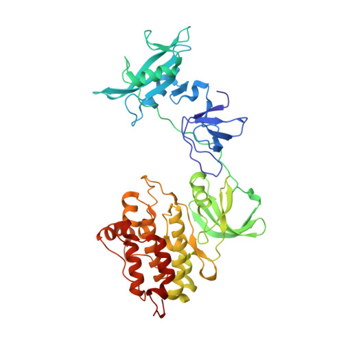
    The regulation of the activity of Abl and Src family tyrosine kinases is mediated by intramolecular interactions between the SH3, SH2, and kinase (SH1) domains. We have determined the crystal structure of an unphosphorylated form of c-Src in which the SH2 domain is not bound to the C-terminal tail. This results in an open structure where the kinase domain adopts an active conformation and the C terminus binds within a hydrophobic pocket in the C-terminal lobe. NMR binding studies support the hypothesis that an N-terminal myristate could bind in this pocket, as observed for Abl, suggesting that c-Src may also be regulated by myristate binding. In addition, the structure contains a des-methyl analog of the antileukemia drug imatinib (STI571; Gleevec). This structure reveals why the drug shows a low affinity for active kinase conformations, contributing to its excellent kinase selectivity profile.


  • Organizational Affiliation

    Discovery Technologies, Novartis Institutes for Biomedical Research, CH-4056 Basel, Switzerland. sandra.jacob@novartis.com


Macromolecules
Find similar proteins by:  (by identity cutoff)  |  3D Structure
Entity ID: 1
MoleculeChains Sequence LengthOrganismDetailsImage
Proto-oncogene tyrosine-protein kinase Src452Homo sapiensMutation(s): 0 
Gene Names: SRC
EC: 2.7.1.112
UniProt & NIH Common Fund Data Resources
Find proteins for P12931 (Homo sapiens)
Explore P12931 
Go to UniProtKB:  P12931
PHAROS:  P12931
GTEx:  ENSG00000197122 
Entity Groups  
Sequence Clusters30% Identity50% Identity70% Identity90% Identity95% Identity100% Identity
UniProt GroupP12931
Sequence Annotations
Expand
  • Reference Sequence
Small Molecules
Binding Affinity Annotations 
IDSourceBinding Affinity
MPZ PDBBind:  1Y57 IC50: 1633 (nM) from 1 assay(s)
Binding MOAD:  1Y57 IC50: 1633 (nM) from 1 assay(s)
BindingDB:  1Y57 IC50: 7800 (nM) from 1 assay(s)
Experimental Data & Validation

Experimental Data

  • Method: X-RAY DIFFRACTION
  • Resolution: 1.91 Å
  • R-Value Free: 0.213 
  • R-Value Work: 0.188 
  • R-Value Observed: 0.189 
  • Space Group: P 41 21 2
Unit Cell:
Length ( Å )Angle ( ˚ )
a = 106.212α = 90
b = 106.212β = 90
c = 123.731γ = 90
Software Package:
Software NamePurpose
REFMACrefinement
DENZOdata reduction
SCALEPACKdata scaling
MOLREPphasing

Structure Validation

View Full Validation Report


Ligand Structure Quality Assessment 


Entry History 

Deposition Data

Revision History  (Full details and data files)

  • Version 1.0: 2005-06-21
    Type: Initial release
  • Version 1.1: 2008-04-30
    Changes: Version format compliance
  • Version 1.2: 2011-07-13
    Changes: Advisory, Version format compliance
  • Version 1.3: 2015-04-15
    Changes: Database references
  • Version 1.4: 2024-03-13
    Changes: Data collection, Database references, Derived calculations