1XHM

The Crystal Structure of a Biologically Active Peptide (SIGK) Bound to a G Protein Beta:Gamma Heterodimer


Experimental Data Snapshot

  • Method: X-RAY DIFFRACTION
  • Resolution: 2.70 Å
  • R-Value Free: 0.287 
  • R-Value Work: 0.227 
  • R-Value Observed: 0.227 

wwPDB Validation   3D Report Full Report


This is version 1.3 of the entry. See complete history


Literature

Structural and Molecular Characterization of a Preferred Protein Interaction Surface on G Protein betagamma Subunits.

Davis, T.L.Bonacci, T.M.Sprang, S.R.Smrcka, A.V.

(2005) Biochemistry 44: 10593-10604

  • DOI: https://doi.org/10.1021/bi050655i
  • Primary Citation of Related Structures:  
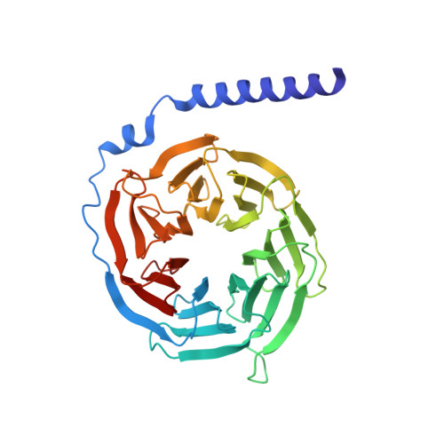
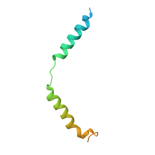
    1XHM

  • PubMed Abstract: 

    G protein betagamma subunits associate with many binding partners in cellular signaling cascades. In previous work, we used random-peptide phage display screening to identify a diverse family of peptides that bound to a common surface on Gbetagamma subunits and blocked a subset of Gbetagamma effectors. Later studies showed that one of the peptides caused G protein activation through a novel Gbetagamma-dependent, nucleotide exchange-independent mechanism. Here we report the X-ray crystal structure of Gbeta(1)gamma(2) bound to this peptide, SIGK (SIGKAFKILGYPDYD), at 2.7 A resolution. SIGK forms a helical structure that binds the same face of Gbeta(1) as the switch II region of Galpha. The interaction interface can be subdivided into polar and nonpolar interfaces that together contain a mixture of binding determinants that may be responsible for the ability of this surface to recognize multiple protein partners. Systematic mutagenic analysis of the peptide-Gbeta(1) interface indicates that distinct sets of amino acids within this interface are required for binding of different peptides. Among these unique amino acid interactions, specific electrostatic binding contacts within the polar interface are required for peptide-mediated subunit dissociation. The data provide a mechanistic basis for multiple target recognition by Gbetagamma subunits with diverse functional interactions within a common interface and suggest that pharmacological targeting of distinct regions within this interface could allow for selective manipulation of Gbetagamma-dependent signaling pathways.


  • Organizational Affiliation

    Department of Biochemistry, University of Texas Southwestern Medical Center, 5323 Harry Hines Boulevard, MC 9050, Dallas, Texas 75390-9050, USA.


Macromolecules
Find similar proteins by:  (by identity cutoff)  |  3D Structure
Entity ID: 1
MoleculeChains Sequence LengthOrganismDetailsImage
Guanine nucleotide-binding protein G(I)/G(S)/G(T) beta subunit 1340Bos taurusMutation(s): 0 
Gene Names: GNB1
UniProt
Find proteins for P62871 (Bos taurus)
Explore P62871 
Go to UniProtKB:  P62871
Entity Groups  
Sequence Clusters30% Identity50% Identity70% Identity90% Identity95% Identity100% Identity
UniProt GroupP62871
Sequence Annotations
Expand
  • Reference Sequence
Find similar proteins by:  (by identity cutoff)  |  3D Structure
Entity ID: 2
MoleculeChains Sequence LengthOrganismDetailsImage
Guanine nucleotide-binding protein G(I)/G(S)/G(O) gamma-2 subunit77Bos taurusMutation(s): 0 
Gene Names: Gng2
UniProt
Find proteins for P63212 (Bos taurus)
Explore P63212 
Go to UniProtKB:  P63212
Entity Groups  
Sequence Clusters30% Identity50% Identity70% Identity90% Identity95% Identity100% Identity
UniProt GroupP63212
Sequence Annotations
Expand
  • Reference Sequence

Find similar proteins by:  Sequence   |   3D Structure  

Entity ID: 3
MoleculeChains Sequence LengthOrganismDetailsImage
SIGK Peptide15N/AMutation(s): 0 
Entity Groups  
Sequence Clusters30% Identity50% Identity70% Identity90% Identity95% Identity100% Identity
Sequence Annotations
Expand
  • Reference Sequence
Experimental Data & Validation

Experimental Data

  • Method: X-RAY DIFFRACTION
  • Resolution: 2.70 Å
  • R-Value Free: 0.287 
  • R-Value Work: 0.227 
  • R-Value Observed: 0.227 
  • Space Group: P 21 21 21
Unit Cell:
Length ( Å )Angle ( ˚ )
a = 45.468α = 90
b = 74.669β = 90
c = 108.023γ = 90
Software Package:
Software NamePurpose
CNSrefinement
HKL-2000data reduction
SCALEPACKdata scaling
PHASERphasing

Structure Validation

View Full Validation Report


Entry History 

Deposition Data

Revision History  (Full details and data files)

  • Version 1.0: 2005-08-09
    Type: Initial release
  • Version 1.1: 2008-04-30
    Changes: Version format compliance
  • Version 1.2: 2011-07-13
    Changes: Version format compliance
  • Version 1.3: 2023-08-23
    Changes: Data collection, Database references, Refinement description