1NXU

CRYSTAL STRUCTURE OF E. COLI HYPOTHETICAL OXIDOREDUCTASE YIAK NORTHEAST STRUCTURAL GENOMICS CONSORTIUM TARGET ER82.


Experimental Data Snapshot

  • Method: X-RAY DIFFRACTION
  • Resolution: 1.80 Å
  • R-Value Free: 0.235 
  • R-Value Work: 0.192 
  • R-Value Observed: 0.192 

wwPDB Validation   3D Report Full Report


This is version 1.2 of the entry. See complete history


Literature

A Novel NAD-binding Protein Revealed by the Crystal Structure of 2,3-Diketo-L-gulonate Reductase (YiaK).

Forouhar, F.Lee, I.Benach, J.Kulkarni, K.Xiao, R.Acton, T.B.Montelione, G.T.Tong, L.

(2004) J Biol Chem 279: 13148-13155

  • DOI: https://doi.org/10.1074/jbc.M313580200
  • Primary Citation of Related Structures:  
    1NXU, 1S20

  • PubMed Abstract: 

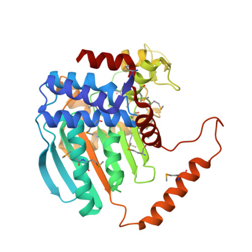
    Escherichia coli YiaK catalyzes the reduction of 2,3-diketo-L-gulonate in the presence of NADH. It belongs to a large family of oxidoreductases that is conserved in archaea, bacteria, and eukaryotes but shows no sequence homology to other proteins. We report here the crystal structures at up to 2.0-A resolution of YiaK alone and in complex with NAD-tartrate. YiaK has a new polypeptide backbone fold and a novel mode of recognizing the NAD cofactor. In addition, NAD is bound in an unusual conformation, at the interface of a dimer of the enzyme. The crystallographic analysis unexpectedly revealed the binding of tartrate in the active site. Enzyme kinetics studies confirm that tartrate and the related D-malate are inhibitors of YiaK. In contrast to most other enzymes where substrate binding produces a more closed conformation, the binding of NAD-tartrate to YiaK produces a more open active site. The free enzyme conformation is incompatible with NAD binding. His(44) is likely the catalytic residue of the enzyme.


  • Organizational Affiliation

    Department of Biological Sciences, Northeast Structural Genomics Consortium, Columbia University, New York, NY 10027, USA.


Macromolecules
Find similar proteins by:  (by identity cutoff)  |  3D Structure
Entity ID: 1
MoleculeChains Sequence LengthOrganismDetailsImage
Hypothetical oxidoreductase yiaK
A, B
333Escherichia coliMutation(s): 15 
Gene Names: YIAK OR B3575
EC: 1.1.1
UniProt
Find proteins for P37672 (Escherichia coli (strain K12))
Explore P37672 
Go to UniProtKB:  P37672
Entity Groups  
Sequence Clusters30% Identity50% Identity70% Identity90% Identity95% Identity100% Identity
UniProt GroupP37672
Sequence Annotations
Expand
  • Reference Sequence
Experimental Data & Validation

Experimental Data

  • Method: X-RAY DIFFRACTION
  • Resolution: 1.80 Å
  • R-Value Free: 0.235 
  • R-Value Work: 0.192 
  • R-Value Observed: 0.192 
  • Space Group: P 1 21 1
Unit Cell:
Length ( Å )Angle ( ˚ )
a = 58.012α = 90
b = 51.229β = 103.78
c = 108.93γ = 90
Software Package:
Software NamePurpose
CNSrefinement
DENZOdata reduction
SCALEPACKdata scaling
SOLVEphasing

Structure Validation

View Full Validation Report


Entry History 

Revision History  (Full details and data files)

  • Version 1.0: 2003-03-11
    Type: Initial release
  • Version 1.1: 2008-04-29
    Changes: Version format compliance
  • Version 1.2: 2011-07-13
    Changes: Version format compliance