The SufE sulfur-acceptor protein contains a conserved core structure that mediates interdomain interactions in a variety of redox protein complexes
Goldsmith-Fischman, S., Kuzin, A., Edstrom, W.C., Benach, J., Shastry, R., Xiao, R., Acton, T.B., Honig, B., Montelione, G.T., Hunt, J.F.(2004) J Mol Biol 344: 549-565
- PubMed: 15522304
- DOI: https://doi.org/10.1016/j.jmb.2004.08.074
- Primary Citation of Related Structures:
1MZG - PubMed Abstract:
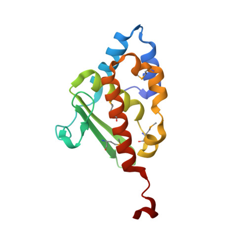
The isc and suf operons in Escherichia coli represent alternative genetic systems optimized to mediate the essential metabolic process of iron-sulfur cluster (Fe-S) assembly under basal or oxidative-stress conditions, respectively. Some of the proteins in these two operons share strong sequence homology, e.g. the cysteine desulfurases IscS and SufS, and presumably play the same role in the oxygen-sensitive assembly process. However, other proteins in these operons share no significant homology and occur in a mutually exclusive manner in Fe-S assembly operons in other organisms (e.g. IscU and SufE). These latter proteins presumably play distinct roles adapted to the different assembly mechanisms used by the two systems. IscU has three invariant cysteine residues that function as a template for Fe-S assembly while accepting a sulfur atom from IscS. SufE, in contrast, does not function as an Fe-S assembly template but has been suggested to function as a shuttle protein that uses a persulfide linkage to a single invariant cysteine residue to transfer a sulfur atom from SufS to an alternative Fe-S assembly template. Here, we present and analyze the 2.0A crystal structure of E.coli SufE. The structure shows that the persulfide-forming cysteine occurs at the tip of a loop with elevated B-factors, where its side-chain is buried from solvent exposure in a hydrophobic cavity located beneath a highly conserved surface. Despite the lack of sequence homology, the core of SufE shows strong structural similarity to IscU, and the sulfur-acceptor site in SufE coincides with the location of the cysteine residues mediating Fe-S cluster assembly in IscU. Thus, a conserved core structure is implicated in mediating the interactions of both SufE and IscU with the mutually homologous cysteine desulfurase enzymes present in their respective operons. A similar core structure is observed in a domain found in a variety of Fe-S cluster containing flavoenzymes including xanthine dehydrogenase, where it also mediates interdomain interactions. Therefore, the core fold of SufE/IscU has been adapted to mediate interdomain interactions in diverse redox protein systems in the course of evolution.
Organizational Affiliation:
Department of Biochemistry and Molecular Biophysics, Howard Hughes Medical Institute, Columbia University College of Physicians and Surgeons, New York, NY 10032, USA.