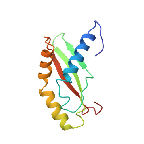
Solution NMR Structure of Ribosome-binding Factor A (RbfA), A Cold-shock Adaptation Protein from Escherichia coli
Huang, Y.J., Swapna, G.V.T., Rajan, P.K., Ke, H., Xia, B., Shukla, K., Inouye, M., Montelione, G.T.(2003) J Mol Biol 327: 521-536
- PubMed: 12628255
- DOI: https://doi.org/10.1016/s0022-2836(03)00061-5
- Primary Citation of Related Structures:
1KKG - PubMed Abstract:
Ribosome-binding factor A (RbfA) from Escherichia coli is a cold-shock adaptation protein. It is essential for efficient processing of 16S rRNA and is suspected to interact with the 5'-terminal helix (helix I) of 16S rRNA. RbfA is a member of a large family of small proteins found in most bacterial organisms, making it an important target for structural proteomics. Here, we describe the three-dimensional structure of RbfADelta25, a 108 residue construct with 25 residues removed from the carboxyl terminus of full-length RbfA, determined in solution at pH 5.0 by heteronuclear NMR methods. The structure determination was carried out using largely automated methods for determining resonance assignments, interpreting nuclear Overhauser effect (NOE) spectroscopy (NOESY) spectra, and structure generation. RbfADelta25 has an alpha+beta fold containing three helices and three beta-strands, alpha1-beta1-beta2-alpha2-alpha3-beta3. The structure has type-II KH-domain fold topology, related to conserved KH sequence family proteins whose betaalphaalphabeta subunits are characterized by a helix-turn-helix motif with sequence signature GxxG at the turn. In RbfA, this betaalphaalphabeta subunit is characterized by a helix-kink-helix motif in which the GxxG sequence is replaced by a conserved AxG sequence, including a strongly conserved Ala residue at position 75 forming an interhelical kink. The electrostatic field distribution about RbfADelta25 is bipolar; one side of the molecule is strongly negative and the opposite face has a strong positive electrostatic field. A "dynamic hot spot" of RbfADelta25 has been identified in the vicinity of a beta-bulge at strongly conserved residue Ser39 by 15N R(1), R(2) relaxation rate and heteronuclear 15N-1H NOE measurements. Analyses of these distributions of electrostatic field and internal dynamics, together with evolutionary implications of fold and sequence conservation, suggest that RbfA is indeed a nucleic acid-binding protein, and identify a potential RNA-binding site in or around the conserved polypeptide segment Ser76-Asp100 corresponding to the alpha3-loop-beta3 helix-loop-strand structure. While the structure of RbfADelta25 is most similar to that of the KH domain of the E.coli Era GTPase, its electrostatic field distribution is most similar to the KH1 domain of the NusA protein from Thermotoga maritima, another cold-shock associated RNA-binding protein. Both RbfA and NusA are regulated in the same E.coli operon. Structural and functional similarities between RbfA, NusA, and other bacterial type II KH domains suggest previously unsuspected evolutionary relationships between these cold-shock associated proteins.
Organizational Affiliation:
Department of Molecular Biology and Biochemistry, Center for Advanced Biotechnology and Medicine, Rutgers University, Piscataway, NJ 08854, USA.