1J91

Crystal structure of Z. mays CK2 kinase alpha subunit in complex with the ATP-competitive inhibitor 4,5,6,7-tetrabromobenzotriazole


Experimental Data Snapshot

  • Method: X-RAY DIFFRACTION
  • Resolution: 2.22 Å
  • R-Value Free: 0.268 
  • R-Value Work: 0.205 

wwPDB Validation   3D Report Full Report


This is version 1.3 of the entry. See complete history


Literature

Structural features underlying selective inhibition of protein kinase CK2 by ATP site-directed tetrabromo-2-benzotriazole.

Battistutta, R.De Moliner, E.Sarno, S.Zanotti, G.Pinna, L.A.

(2001) Protein Sci 10: 2200-2206

  • DOI: https://doi.org/10.1110/ps.19601
  • Primary Citation of Related Structures:  
    1J91, 1JAM

  • PubMed Abstract: 

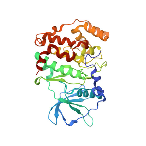
    Two novel crystal structures of Zea mays protein kinase CK2alpha catalytic subunit, one in complex with the specific inhibitor 4,5,6,7-tetrabromobenzotriazole (TBB) and another in the apo-form, were solved at 2.2 A resolution. These structures were compared with those of the enzyme in presence of ATP and GTP (the natural cosubstrates) and the inhibitor emodin. Interaction of TBB with the active site of CK2alpha is mainly due to van der Waals contacts, with the ligand fitting almost perfectly the cavity. One nitrogen of the five-membered ring interacts with two charged residues, Glu 81 and Lys 68, in the depth of the cavity, through two water molecules. These are buried in the active site and are also generally found in the structures of CK2alpha enzyme analyzed so far, with the exception of the complex with emodin. In the N-terminal lobe, the position of helix alphaC is particularly well preserved in all the structures examined; the Gly-rich loop is displaced from the intermediate position it has in the apo-form and in the presence of the natural cosubstrates (ATP/GTP) to either an upper (with TBB) or a lower position (with emodin). The selectivity of TBB for CK2 appears to be mainly dictated by the reduced size of the active site which in most other protein kinases is too large for making stable interactions with this inhibitor.


  • Organizational Affiliation

    Department of Organic Chemistry and CNR Biopolymer Research Center, University of Padova, 35131 Padova, Italy.


Macromolecules
Find similar proteins by:  (by identity cutoff)  |  3D Structure
Entity ID: 1
MoleculeChains Sequence LengthOrganismDetailsImage
CASEIN KINASE II, ALPHA CHAIN
A, B
332Zea maysMutation(s): 0 
EC: 2.7.1.37
UniProt
Find proteins for P28523 (Zea mays)
Explore P28523 
Go to UniProtKB:  P28523
Entity Groups  
Sequence Clusters30% Identity50% Identity70% Identity90% Identity95% Identity100% Identity
UniProt GroupP28523
Sequence Annotations
Expand
  • Reference Sequence
Small Molecules
Ligands 1 Unique
IDChains Name / Formula / InChI Key2D Diagram3D Interactions
TBS
Query on TBS

Download Ideal Coordinates CCD File 
C [auth A],
D [auth B]
4,5,6,7-TETRABROMOBENZOTRIAZOLE
C6 H Br4 N3
OMZYUVOATZSGJY-UHFFFAOYSA-N
Experimental Data & Validation

Experimental Data

  • Method: X-RAY DIFFRACTION
  • Resolution: 2.22 Å
  • R-Value Free: 0.268 
  • R-Value Work: 0.205 
  • Space Group: P 1
Unit Cell:
Length ( Å )Angle ( ˚ )
a = 48.255α = 89.77
b = 55.943β = 102.51
c = 60.302γ = 99.27
Software Package:
Software NamePurpose
AMoREphasing
CNSrefinement
MOSFLMdata reduction
CCP4data scaling

Structure Validation

View Full Validation Report


Entry History 

Deposition Data

Revision History  (Full details and data files)

  • Version 1.0: 2002-05-29
    Type: Initial release
  • Version 1.1: 2008-04-27
    Changes: Version format compliance
  • Version 1.2: 2011-07-13
    Changes: Version format compliance
  • Version 1.3: 2024-02-07
    Changes: Data collection, Database references, Derived calculations