Glutamate 170 of human l-3-hydroxyacyl-CoA dehydrogenase is required for proper orientation of the catalytic histidine and structural integrity of the enzyme.
Barycki, J.J., O'Brien, L.K., Strauss, A.W., Banaszak, L.J.(2001) J Biol Chem 276: 36718-36726
- PubMed: 11451959
- DOI: https://doi.org/10.1074/jbc.M104839200
- Primary Citation of Related Structures:
1IL0 - PubMed Abstract:
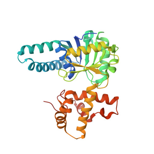
l-3-Hydroxyacyl-CoA dehydrogenase (HAD), the penultimate enzyme in the beta-oxidation spiral, reversibly catalyzes the conversion of l-3-hydroxyacyl-CoA to the corresponding 3-ketoacyl-CoA. Similar to other dehydrogenases, HAD contains a general acid/base, His(158), which is within hydrogen bond distance of a carboxylate, Glu(170). To investigate its function in this catalytic dyad, Glu(170) was replaced with glutamine (E170Q), and the mutant enzyme was characterized. Whereas substrate and cofactor binding were unaffected by the mutation, E170Q exhibited diminished catalytic activity. Protonation of the catalytic histidine did not restore wild-type activity, indicating that modulation of the pK(a) of His(158) is not the sole function of Glu(170). The pH profile of charge transfer complex formation, an independent indicator of active site integrity, was unaltered by the amino acid substitution, but the intensity of the charge transfer band was diminished. This observation, coupled with significantly reduced enzymatic stability of the E170Q mutant, implicates Glu(170) in maintenance of active site architecture. Examination of the crystal structure of E170Q in complex with NAD(+) and acetoacetyl-CoA (R = 21.9%, R(free) = 27.6%, 2.2 A) reveals that Gln(170) no longer hydrogen bonds to the side chain of His(158). Instead, the imidazole ring is nearly perpendicular to its placement in the comparable native complex and no longer positioned for efficient catalysis.
Organizational Affiliation:
Department of Biochemistry, Molecular Biology, and Biophysics, University of Minnesota, Minneapolis, Minnesota 55455, USA.